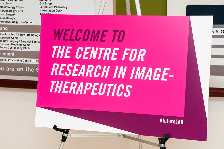
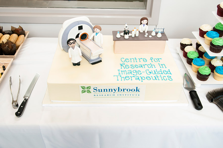
The FutureLab is the $160-million Centre for Research in Image-Guided Therapeutics at Sunnybrook Research Institute (SRI), and it is unique in Canada.
In this centre, scientists are developing, testing and refining basic science and medical imaging technologies and therapeutics—including new drugs, biological agents and imaging devices—and translating them into the clinic for the benefit of patients. Building the centre has added 150,000 square feet of world-class facilities to SRI, including:
The centre was established by a $75-million grant from the Canada Foundation for Innovation, with investment from industry and other funding partners.
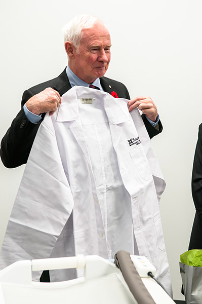
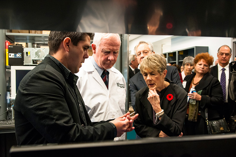
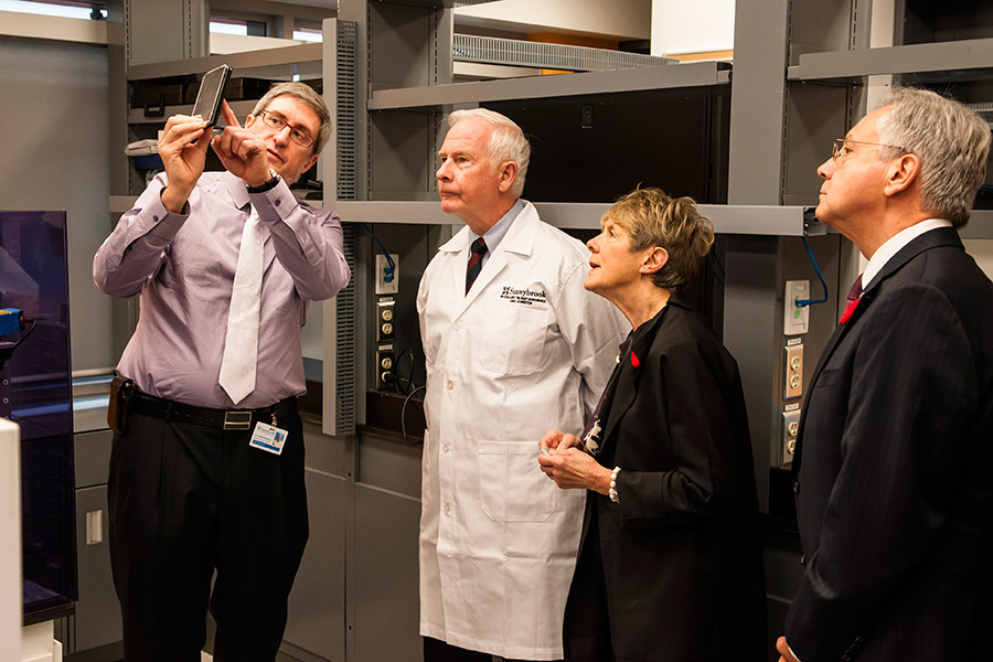
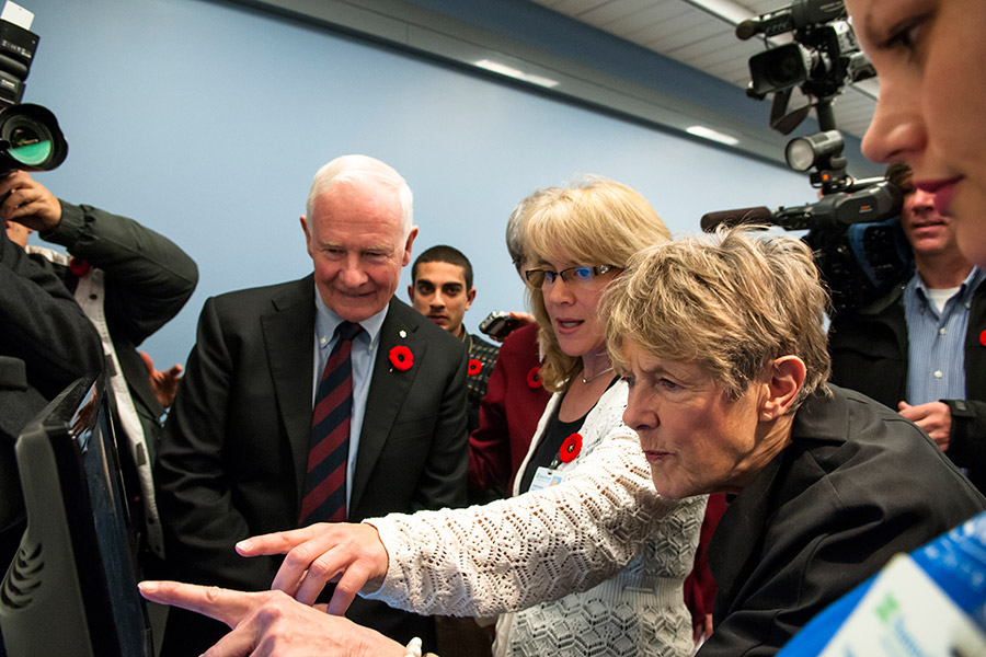
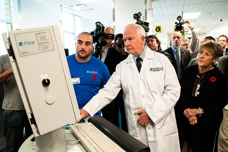
To celebrate the official launch, we invite you to tour our labs and experience the science of the centre.
This lab brings together all Sunnybrook Research Institute (SRI) scientists and engineers working to analyze and develop new image-processing methods for cancer, and heart and brain conditions. Visitors will be able to “diagnose” disease using advanced image-processing software, for example, to do brain image segmentation. They can try their hand at Vurtigo, a 4-D, real-time visualization software program for guiding cardiovascular interventions that was developed here at SRI. It is designed to be used with a magnetic resonance imaging (MRI) scanner, actively tracked catheters and navigational devices. They can also pan, rotate and zoom through a 3-D dataset of cardiac images using a six-degrees-of-freedom mouse—which gives the effect of reaching into the screen and holding the image in hand.
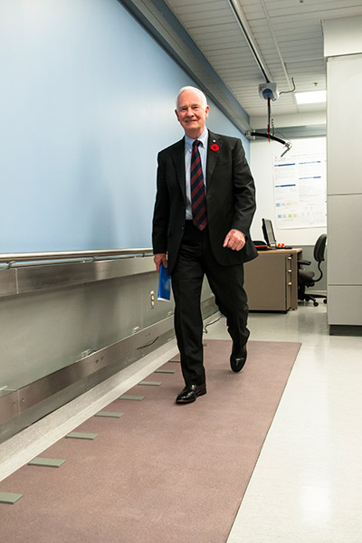
The neurointervention centre is developing better ways to treat brain disorders like stroke and dementia, and to help people recover after stroke. Here, visitors can assess their mobility by walking on the gait analysis mat. Visitors will see how fast they are walking, how far apart their feet are and the force exerted on different parts of their feet. They can also up the ante by doing a brain test while walking to see how that affects their gait. This equipment is used to track a patient’s recovery after stroke. Visitors will also get to test their balance control, using force plates, which will give them information about their body’s symmetry, and the Wii balance board. A demonstration of the XSENS system, a set of wireless sensors that attach to a person, will show visitors how researchers are using the 3-D system to track human motion in real time.
Visitors can also learn how one researcher is studying the effects of exercise on the brain; experience it feels like to be in an magnetic resonance imaging (MRI) scanner with the MRI simulator; see how researchers are using image-guided stimulation and TMS to stimulate the brain noninvasively; and try a variety of cognitive assessments.
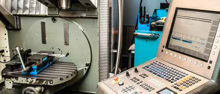
In our advanced machine shop, part of the device development lab, visitors can participate in demos of state-of-the-art device fabrication machines, including a two-ton waterjet, which is a machine that can cut through a substance as hard as titanium or as soft as a fig Newton without heating or warping it. It can make any 3-D pattern and etch fine detail. They will also see the shop’s computer numerical control milling machine and lathe in use. These machines use computer automated design software to cut parts precisely for prototype devices and have microfabrication capabilities. There will be a table displaying clinical and research items made in the machine shop that visitors can examine. At another station, visitors will see a 3-D rapid prototyper in action—a machine that is used to sculpt medical parts using computer-aided design. These machines are used to craft components for the devices we are making, such as an ultrasound probe that is integral to a new technology to treat prostate cancer with focused ultrasound—a therapy an SRI scientist developed and commercialized. We are the only research institute in Canada with such a lab.
In the orthopaedic biomechanics lab, visitors will see how bioengineering research is aimed at maximizing function among individuals who develop musculoskeletal disease or disability. Here, visitors will look at craniofacial bones on imaging systems to understand how researchers look at clinical CT scans to detect fractures. Also on display, will be a prototype device invented by an SRI scientist and colleagues. The “bone tape” is like duct tape for the face—it works to adhere fractured facial bones under the skin, to speed the healing process. At another station, visitors can put screws in the spine using the pedicle screw simulator and look at CT scans of spinal metastases on a computer system to learn how researchers quantify bone risk fracture. Visitors will also see an O-arm surgical imaging system, which is used to develop minimally invasive procedures for musculoskeletal surgery.
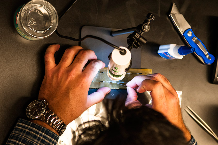
In the device fabrication room, visitors will see machining tools such as a mill, lathe, dicing saw and micro drill in use. They will learn about how this equipment is used to make hair-thin cuts—as fine as 15 microns, about the width of a strand of hair—and small holes for parts such as connectors and adapters that are used in ultrasound probes and catheters. They will also see a lapping system that uses aluminum oxide to grind material finely. This machine is used to produce sensors for low- and high-frequency ultrasound devices, including the 3-D intravascular ultrasound catheter invented by an SRI scientist. Visitors will see samples of these components on display. They can use a micrometre, a staple of a micromachine shop that measures to one-thousandth of a millimetre, to measure extremely thin objects, like sheets of paper.
At this station, visitors can see the focused ultrasound brain system that consists of a patient table and a helmet-like device, and that is the central component of the technology that Time Magazine named as a top 50 invention in 2011. The scientist who invented this technology is the head of CeRIGT. We recruited him from Harvard five years ago. InSightec, an Israeli company, commercialized the technology. It enables the destruction and removal of tumours and other lesions deep inside the brain and body without cutting through skin. Visitors will learn how this world-changing technology focuses many ultrasound beams through the skull, into the brain, onto a precise “hot spot”—the therapeutic target, under guidance by magnetic resonance imaging. This revolutionary “surgery” is being tested in clinical trials, some of which are Canadian firsts, including one to destroy inoperable brain tumours. Visitors will also learn how the device is being further developed to enable delivery of drugs and other biological agents across the blood-brain barrier, for example to treat Alzheimer’s disease.
Several CeRIGT researchers are designing and testing cardiac devices for use with imaging technology. In the catheter testing lab, visitors will learn how SRI scientists and their teams build catheters by using specialized equipment that measures specific properties such as torque and puncture force. They will operate this equipment and test the “pushability” of a catheter by moving it through a simulated vessel and retracting it, while the system calculates the catheter’s position and the resistance encountered as it moves through bends in the vessel. Visitors will also see how components for catheters are made at the hot air work station. They will see a demonstration of this equipment, which is used to shape parts through the precise application of extremely hot air. They will get to use it by activating the cold air function, used to set the molded material.
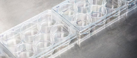
Here, visitors will learn about how one CeRIGT researcher is developing cell-based therapies to help burn patients. At the tissue culture hood, visitors will use a pipette gun to change liquid media on a six-well plate. They will learn how the lab obtains and cultures stem cells. They will also learn about the lab’s methods of studying and preparing these cells for its wound-healing projects.
At the cell culture microscope, visitors will play “cell detective” as they study cells on a plate to distinguish liver cells from stem cells that can differentiate into other cell types. They will learn how staining protocols are used to observe the differentiation of stem cells into bone, cartilage and fat cells. There will also be a video describing the lab’s collaboration with a U of T researcher to make a “skin printer,” technology that may one day be used to make customized skin to repair major burns and other wounds. Visitors will also learn about the group’s work to develop biological scaffolds— inexpensive synthetic skin using polymers and stem cells, to replace donor skin grafts. The group has made prototypes and confirmed that stem cells can survive in the scaffold.
Visitors to the this space will see the Opera system—one of only four in the world—in use. It is the first fully automated high-content screening microscope that can image fluorescence lifetime, the property most of interest to drug companies, because it tells them if a drug is working. This system can take 100,000 pictures a day without anyone being there! Visitors will see the system’s robotic arm load biological samples into plates for imaging and analysis, and learn how the system is used to study the proteins involved in cell death—vital information in the high-risk, high-reward domain of drug discovery. At the Work Cell station, visitors will see another high-tech cell analysis machine at work and meet “Val,” the robotic arm that delivers plates of cells to platforms within the system. This system automates the arduous task of depositing cells into the tiny cell wells used by biologists, and transfers molecules to cells systematically in a sterile environment. Visitors can try pipetting by hand, and then see how much faster the automated system is. They can also look through a microscope to watch proteins tagged with a light-emitting material interact.
In this chemistry lab, visitors will learn how scientists are engineering biomolecules to develop therapies for cancer. Visitors will use a Nanodrop spectrophotometer that tests nano-sized—super small—samples for the presence of protein or DNA. At the ultraviolet box station, visitors will learn about the lab’s bacterial toxin research. Here, they will learn how to detect deadly bacteria on a contaminated object. They will swab rubber steaks, one contaminated, one not, and then place their swabs under ultraviolet light to identify which one is contaminated. At the molecular design station, visitors will see how the lab redesigns biomolecules by modifying their structures. Visitors will “handle” the structure of a molecule displayed on a screen. They will work at the submicron scale to identify relevant areas for the reengineering of a new derivative molecule. One example is a toxin fragment that they reengineered for drug delivery. At the xCelligence station, visitors will look at the kinetics of cell dynamics and learn about how the lab used an emerging technology to devise a way of evaluating the effectiveness of a breast cancer vaccine it developed, and that is poised for clinical trials at the Odette Cancer Centre.
In our stem cell biology lab, visitors can slip on lab coats and become immunologists. Through a series of hands-on stations, visitors can perform the main tasks of a biologist. Through a stepwise process they will learn how to “purify” T cells and work under a tissue culture hood, a sterile environment where cells can be manipulated safely. They will be shown the principles behind magnetic cell sorting. They will perform real cell biology techniques, like staining proteins to look for a specific gene. We will also have a liquid nitrogen station, where visitors can freeze and then smash flowers, thereby demonstrating cryopreservation, a main method of the cell biologist. The aim of this research is to find ways to rebuild a devastated immune system from the inside out. The scientist leading this work is world-renowned for inventing a method to grow an unlimited number of T cells in the lab, and his research has many potential clinical applications. Scientists in this centre are working to “program” T cells to look for and destroy cancer cells by identifying markers and receptors on tumour cells.
This is the wet lab associated with the cell-killing robot room. This lab is manipulating protein-protein interactions to develop ways to control cell death—to hasten it for cancerous cells, and to halt the death of healthy brain cells, for example, in stroke. At the wet benches in this space, visitors can “clone” DNA in bacteria to extract a specific protein for further study, and complete the steps involved in protein purification.
In this space, visitors can see a slide show on cardiac tissue characterization, to learn why this is important in designing better ways to diagnose, predict the outcome of and treat heart attack. They will see images of damage to the heart muscle following a heart attack and learn how SRI scientists are using magnetic resonance imaging (MRI) to understand what is happening in these patients, toward guiding therapeutic approaches.
Down the hall, visitors will learn about the devices engineered by an SRI clinician-scientist-entrepreneur to improve the safety and success rates of cardiac procedures. At the ultrasound catheter station, visitors will see a real-time imaging demonstration of a catheter he invented and commercialized, which uses ultrasound imaging to take high-resolution 3-D images inside the heart to guide minimally invasive procedures. Visitors will also see a prototype of the ergonomic patient bed enhancement he designed to enable cardiologists to perform interventions more comfortably and conveniently in cath labs. Visitors will also see how the lab uses computer-aided design to craft custom anatomical parts using 3-D technology. These life-size parts, which will be on display, are used to test medical devices invented in the lab. An example is the bespoke catheters he will build to treat patients with rare anatomic abnormalities.
In the adjacent bay, visitors will see how one scientist is inventing ways to detect and image disease processes at the molecular level using MRI. At the catheter assembly station, visitors will use a digital camera microscope to assemble the tiny parts used to make a novel catheter. The catheter was invented by an SRI scientist to solve the problem of how to see inside blocked blood vessels in the heart while the patient is in an MR scanner. This station will offer an exclusive glimpse of a technology in development, before it becomes a standard tool in the cardiac surgeon’s kit. While the main focus of this lab’s work is cardiac, they are also doing work that applies to breast cancer. Thus, visitors will learn how to tune an MRI coil by passing their hands in front of a network analyzer and, in effect, becoming part of the electromagnetic circuit.
Visitors to this core facility within CeRIGT will see how it enables visualization and analysis of cells for any biological application. In the analyzer room, visitors can peer inside a 14-parameter cell analyzer and see the optical pathways used to excite and collect the emission of light from cells tagged with a fluorescing agent. They will also see a clinical analyzer in use as it processes thousands of cells from a blood sample. This equipment is connected to a computer, where data such as antigens on cell surfaces (substances that cause the immune system to create antibodies against them) are shown on screen and stored for future use. At the magnetic cell separator, visitors will see T cells labelled with iron particles sorted through magnetic columns.
At the flow sorter station, visitors can look inside a machine’s chamber and see an electrostatic field direct cells into collection vessels. Visitors can activate a flow sort and assign cells to be sorted into culture plates. In the microscopy room, visitors can look at a kidney specimen through a microscope, and optical slices of the hair cells lining the inner ear, and learn about research aimed at restoring hearing and balance.
In the focused ultrasound development lab, researchers are developing high-power, image-guided ultrasound phased-array technology, including transducer arrays and electronics. Here, visitors will see a laser vibrometer, which uses the Doppler effect to make real-time maps of the surface vibrations of ultrasound transducers. This allows researchers to characterize the performance and refine the designs of imaging and therapy transducers.
Visitors will also see the high-power therapy transducer (fountain). At the core of high-intensity focused ultrasound (HIFU) technology—including that showcased in the Focused Ultrasound Brain System station—is the ultrasound transducer. Unlike the low-intensity imaging transducers used in clinics globally, these HIFU transducers are designed to deliver large amounts of power to a small and precisely positioned location in the body, so that ultrasound treatments can be localized to affect only the desired region. The HIFU is paired with magnetic resonance imaging (MRI) to help achieve this precision. An ultrasound fountain demonstrates the energy delivered via focused ultrasound. Visitors will see how we use an ultrasound fountain in our experiments to locate the focus of the ultrasound transducer in an MRI scanner for targeted treatment.
In addition, visitors will see the transcranial therapy and monitoring array. The skull is a significant barrier to ultrasound treatment of the brain. One way to mitigate its effect is to use many ultrasound elements positioned over a large area, a technique this lab is pioneering. With integrated ultrasound receivers, this dome-shaped array is helping researchers refine techniques for ultrasound treatment and monitoring through the skull. Potential treatment implications, such as for brain cancer or Alzheimer's disease, are staggering.
Here, visitors will see how researchers are using 3-D histopathology techniques to advance research in cancer and diseases of the brain and heart. In the whole-mount processing lab, visitors will see a demonstration on serial, whole-mount sectioning and a display of paraffin blocks (before sectioning) and glass slides (after sectioning) that show the contrast between conventional sampling-based and 3-D pathology for a variety of tumour sites.
Using the GE multiplexer, visitors can sleuth for biomarkers. This unique device allows researchers to look at multiple areas where a particular biological marker is present. Visitors will then look at slides through a microscope to compare with single biomarker expression.
In the image digitization room, visitors will see a presentation of a tumour using whole-mount sections and 3-D visualization and its correlation with other ex-vivo imaging. Visitors will also get to look through a microscope and compare glass slides from selected cases.
In addition, visitors will learn about genome sequencing. Researchers are using DNA sequencing to identify which patient tumours will or will not benefit from a particular drug therapy. This technology will be used more widely clinically as its costs fall and understanding how to use the information grows. Here, visitors will learn about validation technologies, and see DNA mapping of sequencing runs with and without mutation and RNA mapping of expression levels.