Hybrid XMR system
An increasing number of treatments for cardiac and vascular diseases is performed using minimally invasive techniques. Access to the treatment site is achieved via a small puncture in a blood vessel located under the skin that allows the insertion of a catheter. Blood vessels provide access to nearly all organ systems and this method of treatment delivery is generally less invasive than surgery and allows for faster patient recovery. Minimally invasive techniques can be the only option for patients who cannot tolerate surgery.
The physician cannot directly see the treatment site and must rely on medical imaging techniques such as X-ray and MRI to evaluate the progress of the procedure deep inside the patient. X-ray images can be acquired at rates up to 30 frames per second with a resolution of 0.15 mm providing excellent treatment device guidance in real-time. However, these images are two-dimensional projections providing no depth information and lack the ability to distinguish soft tissues. Figure 1 shows a novel aortic valve designed to be inserted and deployed using a catheter. The procedure requires real-time guidance provided by X-rays and three-dimensional imaging capabilities and soft tissue discrimination provided by MRI to ensure proper positioning.
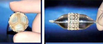
Figure 1: An aortic valve designed for insertion via a catheter. R. Beekman, Congenit. Heart Dis., vol. 1, pp. 2-9, ©2006.
The IRRCI is developing an X-ray imaging system in collaboration with Stanford University that is MRI-compatible and can be operated within one to two metres from the MRI scanner. The proximity of these systems allows rapid switching between the two imaging modalities. Previous attempts to build hybrid systems with both devices in proximity were achieved by making compromises that impacted negatively on the diagnostic quality of the MRI and X-ray images.
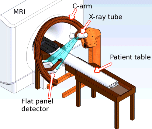
Figure 2: Conceptual drawing of an X-ray C-arm system for use in proximity of a MRI scanner.
Operation of an X-ray system using a high-output rotating anode X-ray tube and flat panel X-ray imaging detector next to the MRI poses many significant challenges which we have now overcome. In an X-ray tube, radiation is produced by accelerating an electron beam in vacuum into a rotating metal target. The electrons experience sudden deceleration in the metal target to produce large amounts of heat and X-rays. The magnetic field bends the electron beam causing misalignment of the X-ray field resulting in the presence of radiation beyond the boundaries of the X-ray detector. This radiation adds dose to the patient with no benefit. The metal target is rotated to prevent the electron beam from hitting the same area of the target for too long causing extreme heating. The magnetic field from the MRI unit reduces the rotation speed of the target requiring the X-ray intensity to be reduced.
We have developed techniques to shield the X-ray tube motor so that it can operate at normal speed, and we have shown that the bending of the electron beam can be corrected by applying a compensating magnetic field using a pair of Helmholtz coils. The X-ray flat panel detector can operate in a magnetic field with some modifications to components such as fan motors. We designed and developed a workstation to control the generation of X-rays and acquisition of images from the system. We are currently designing and building the C-arm allowing the X-ray tube and detector to be positioned to any desired angulations around the patient as is currently possible with a conventional system.
This work is possible due to expertise in electronic and mechanical design within Sunnybrook and collaborations with companies, many of which are in the Toronto area. Sentinelle Medical Inc. (Toronto), a manufacturer of MRI breast biopsy system, is helping the design of the MRI-X-ray compatible patient table. CPI (Georgetown) has provided X-ray generators and expertise to interface their product into our system. Rytech X-ray Inc. (Toronto) has provided us with a custom designed X-ray tube free of ferromagnetic materials used to investigate tube operation in a magnetic field.

