Thinking small


By Alisa Kim
Breakthroughs in science are often the result of creativity and, sometimes, a little serendipity.
Two years ago, Dr. Stuart Foster, an imaging physicist at Sunnybrook Research Institute, was on a bus headed for a tour of the Guinness beer factory in Dublin, Ireland. Sitting next to him was Dr. Mikhail Shapiro, then a postdoc at the University of California at Berkeley. They were in Dublin for the World Molecular Imaging Congress where Shapiro spoke about biologically derived gas nanostructures that could serve as a contrast agent for magnetic resonance imaging (MRI).
“He’d given a paper on use of these particles for MRI, and I was intrigued by it and thought it would be better applied to ultrasound than to MR,” says Foster, who holds the Canada Research Chair in Ultrasound Imaging.
The chance meeting led the researchers to collaborate on a study published in Nature Nanotechnology showing for the first time that the gas nanostructures can be used as molecular “reporters” in ultrasound imaging.
Foster notes that ultrasound needs gas for contrast enhancement and that for the past 45 years, microbubbles, gas bubbles that are injected into the circulation, have been used. Microbubbles can image vascular targets but cannot travel outside blood vessels into tissue. “They’re restricted to a narrow range of applications inside the vessels themselves,” he says. “That there might be a particle that would be useful and be able to get outside the vascular system to report on other targets has been elusive.”
Until now.
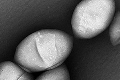
The gas particles grow in bacteria and are harvested via centrifugation, a process used to separate materials suspended in liquid by spinning the liquid at a high speed. The purified gas particles, which are about one-hundredth the size of a red blood cell and invisible to the naked eye, are so tiny that they seep through vessels, yet are detected with ultrasound imaging.
By putting the genes of the gas particle inside other cells and thus “teaching” them to express those genes, researchers could theoretically use the nanostructures to track cancer cells, to study the spread of cancer, for example. The gas particles might also be used one day to trace the activity of stem cells.
“Essentially the particles ‘light the tissue up’ as they circulate and that’s how we follow them,” says Foster, who likens them to green fluorescent protein, found in the jellyfish Aequorea victoria, which glows green under fluorescent light and is used to monitor, among other things, gene expression, protein-protein interactions and cell division in living cells.
That the bacteria developed the gas particles is an evolutionary quirk, says Foster, who notes the gas enables bacteria to float and move closer or farther away from the sun, and to roam to wherever nutrients are present. “You can imagine bacterial particles ‘manufacturing’ thousands of tiny nanostructures that contain gas that give them buoyancy to rise in the liquid soup that they’re living in, and then programming the destruction of those particles when they want to go back down. That’s how they navigate.”
The researchers have used the gas particles in preclinical ultrasound imaging, but much needs to be done before they can be used for human imaging, says Foster. For starters, the surface of the particles needs modification so that they are not attacked by the body’s immune system. They also need to understand the genetic mechanisms to be able to manipulate the particles’ size and shape, which determines how they respond to an ultrasound field.
“We don’t want to use the gas particles for buoyancy; we want them to create the maximum amount of scattered ultrasound from the interaction with the beam. That might be a bit different than what nature intended, but hopefully we can sort those things out.”