


Understanding your heart condition
Our health care team has worked together with previous patients & their experiences to help answer some of the questions that you may have about your upcoming surgery.
The waiting period prior to heart surgery is a very stressful time for patients and their families. Anxiety and emotional changes are normal during this period. It is important to discuss your feelings with family members or friends to help you deal with these emotions.
Knowing how your heart works and what your condition means can help you better understand your surgery and recovery. If you have any questions, please let your care team know. They can provide specific information about your care. You can also contact the cardiovascular coordinators at 416-480-6078.
Your heart
Your heart is a muscle which pumps blood carrying oxygen and nutrients to all parts of the body. It must also provide its own blood supply. The heart is located in the centre of the chest, slightly to the left and is protected by the breastbone (sternum) and ribcage.
The heart contains four chambers:
- Right Atrium
- Right Ventricle
- Left Atrium
- Left Ventricle
Two upper chambers (atria) receive blood from veins. The two lower chambers (ventricles) pump blood out of the heart through arteries. The main pump of the heart is the left ventricle.
The heart also contains four valves:
- Tricuspid Valve (T)
- Mitral Valve (M)
- Pulmonic Valve (P)
- Aortic Valve (A)
In a way, your heart can be compared to four rooms of a house. The valves act as a doorway that only lets blood through in one direction from one part of the heart to the next.
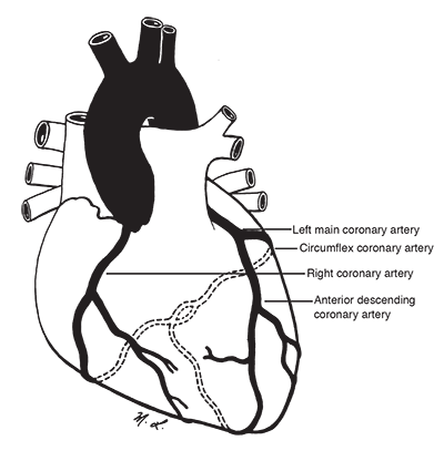
Coronary arteries
The coronary arteries supply blood rich in oxygen to the heart muscle. They lie on the surface of the heart. There are two major coronary arteries.
- The right coronary artery - nourishes the right side and under surface of the heart.
- The left coronary artery - nourishes the left side and the upper surface of the heart. It has a short beginning branch called the left main coronary artery before it divides into two main branches.
The anterior descending coronary artery nourishes the front of the left side of the heart.
The circumflex coronary artery nourishes the back of the left side of the heart.
Coronary artery disease
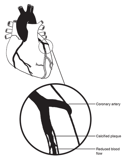
The coronary arteries may be affected by atherosclerosis. (hardening of the arteries). This is a progressive disease where the inner lining of the artery becomes thickened and narrowed with cholesterol and plaque. When this happens there is a decrease in oxygen rich blood carried to the heart muscle.
Angina
When the arteries become partially or completely blocked, by plaque or a blood clot, the blood flow is reduced so much that the cells of the heart may become stressed or permanently damaged. Angina (chest pain) is a warning sign that the heart is stressed from a lack of oxygen. Angina is usually a tight constriction or heaviness in the chest, occasionally an ache or pain in the jaw or arm, or heavy breathing. Blood flow to the heart may be impaired, without angina symptoms, but other evidence may be seen on EKG or with stress testing (silent angina).
Sensations that may indicate angina
- Indigestion
- Fullness
- Numbness or tingling in any part of the arm
- Discomfort in the neck, or back, between the shoulder blades
- Pressure, squeezing, aching, burning, or cramping pain in the chest or neck
- Tightness
- Choking
- Pain in the jaw
- Shortness of breath
Women and men may exhibit very different types of angina pain or symptoms. Not everyone has the same symptoms, and some people have none. For example, many women often describe angina as a sore throat, indigestion, arm heaviness or back pain.
Angina pains can occur when the work of the heart is increased by:
- Exertion
- Exercise
- Excitement
- Anxiety
- Eating a large meal
- Weather (cold or humidity)
When angina cannot be controlled with medical therapy or other non-surgical procedures, bypass surgery may be indicated.
Learn how to reduce your risk factors for coronary artery disease
Valvular heart disease
 Heart valves direct the blood flow through the heart's chambers. Your heart has four valves:
- Tricuspid valve: located between the right atrium and right ventricle
- Mitral valve: located between the left atrium and left ventricle
- Pulmonic valve: located between the right ventricle and pulmonary artery
- Aortic valve: located between the left ventricle and aorta
Valves act like one way doors. They let blood in and out of the heart's chambers each time it beats. Problems are more severe if the defective valves are on the left side of the heart (aortic and mitral valves). These valves regulate blood flow through the main pumping chamber.
Damage to heart valves can occur from:
- Birth defects
- Infection
- Rheumatic fever (or scarlet fever)
- Heart muscle damage
Damage to the heart valves can result in:
- Stenosis (narrowing of the valve opening which makes it difficult to open)
- Insufficiency (the valve does not close properly, also known as regurgitation)
Damage to the valves makes your heart work harder to pump blood to the body and after a number of years may result in heart strain or failure. A damaged heart valve may allow blood to flow in the wrong direction. Although medications may improve the work of the heart and relieve the strain or failure, often surgery to improve the valve disease may be necessary.
