
Tool kit: Olympus multiphoton laser scanning microscope
Sunnybrook Research Institute (SRI) has a new multiphoton microscope. The Olympus FluoView FVMPE-RS multiphoton laser scanning microscope supports brain-related projects at SRI that are working to develop novel therapeutics and improve delivery of existing therapies. It can visualize changes in living cells and tissues that are caused by neurodegenerative diseases, stroke and cancer, and in response to treatments.
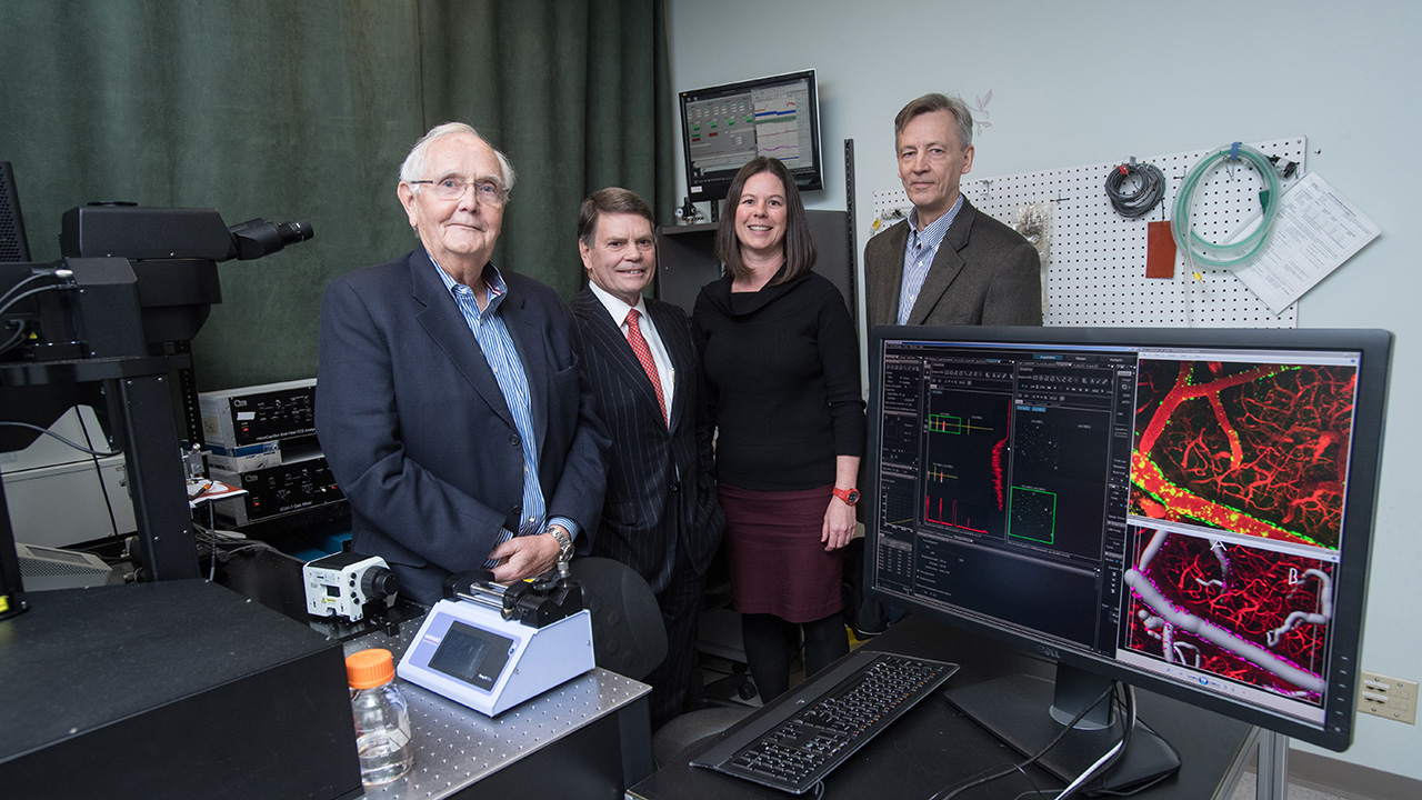
“It is being used in projects aimed at optimizing delivery of therapeutics across the blood-brain barrier via focused ultrasound,” says Dr. Bojana Stefanovic, a senior scientist in the Hurvitz Brain Sciences Research Program, who is a primary user of the system. “Our set-up is unique. We focus on in vivo neuroimaging in preclinical models of disease and optimization of image-guided therapy using focused ultrasound.” Sunnybrook Research Institute leads in this research, and was recently named Canada’s first Centre of Excellence in focused ultrasound.
With its extended infrared range of up to 1300 nm excitation, the system is equipped with high-speed resonant scanners in addition to the conventional galvanometer scanners. This means it can image deep within a specimen. It can image 30 frames per second with 512 x 512 frames; or 438 frames per second with 512 x 32 frames, the fastest commercially available. This high temporal resolution is of particular interest for brain applications, says Stefanovic. Typical imaging sessions take about three hours.
Other SRI researchers who will use the multiphoton microscope include Drs. Isabelle Aubert, David Goertz, Kullervo Hynynen and JoAnne McLaurin. The collective effort is to understand the biological effects of focused ultrasound with and without therapy, and the neurovascular changes associated with progression of amyloid and tau pathologies in Alzheimer’s disease.
The equipment was purchased with the generous support of philanthropists Messrs. Bob Beamish and Gerry Connor. The microscope was installed in December 2016. It will allow researchers to build upon work conducted on an older twin FV1000-MPE system, which is still in operation.