
Discovering how cancer might defy drug therapy
For a cancer cell to die, certain proteins must do their jobs. Given the opportunity, these proteins launch programmed cell death, an essential function that destroys destructive cells and clears out unnecessary ones. Unfortunately, these proteins aren’t always able to kill cancer cells. Dr. David Andrews, director of Biological Sciences at Sunnybrook Research Institute (SRI), aims to find out why, and then figure out how to let them fulfil their duties.
In his latest paper in eLife, Andrews, who is also a professor in the department of biochemistry at the University of Toronto and who holds the Canada Research Chair in membrane biogenesis, has leapt toward achieving that goal. He studies apoptosis [apa-toe-sis], the scientific term for programmed cell death. In the paper, he outlines his lab’s discovery that Bim, a protein essential to apoptosis, binds to anti-cell-death proteins at two sites—not one, as had been thought. This could explain Bim’s role in cancer treatment resistance and why some drugs meant to help with apoptosis fall flat. The finding could also play a part in developing new drugs that permit programmed cell death to kill cancer cells, thus making cancer therapies more effective.
In a cell, there exists a balance between pro-cell-death proteins, like Bim, and anti-cell-death proteins, two of which are Bcl-2 and Bcl-XL. When balanced, the anti-death proteins bind all the pro-death proteins and the cell lives. Most often when a cell becomes cancerous, the pro-death proteins increase; once they’ve outnumbered the anti-death proteins, the cancer cell dies. When a tumour develops, it’s usually because there has been an increase in anti-death proteins, and therefore the call to kill the cancer cells goes unanswered.
About 10 years ago scientists discovered that drugs could be delivered to unlock pro-death proteins from anti-death proteins. For this to happen, drugs mimic the pro-death proteins. This imitation tricks the anti-death proteins into binding to the drugs instead, thereby leaving Bim and other pro-death proteins free to trigger cell death. “When you add a drug, it breaks the balance. The drug binds to anti-death proteins so the pro-death proteins are released and free to function,” says Dr. Qian Liu, a research associate in Andrews’ lab and the first author on the eLife paper.
In other words, this allows treatments, like chemotherapy, to be successful in killing cancer cells.
Scientists noticed, though, that Bim was remaining bound to some of the anti-death proteins, namely Bcl-2 and Bcl-XL, in the presence of drugs. Apoptosis, therefore, couldn’t be induced. In a clinical setting, this continued attachment could render therapy ineffective.
In his study, Andrews and his lab pinpointed just how Bim stays bound to anti-death proteins after drugs were delivered. “What Qian showed is that there is a second binding site between the two proteins, one that’s not near the first binding site,” Andrews says.
This makes Bim the first pro-death protein that has been shown to “double-bolt lock” onto Bcl-XL and Bcl-2, meaning the proteins are secured in two places. Since drugs designed to detach the proteins target only one binding site, this second location prevents the separation of Bim from the anti-death proteins. “The drug cannot compete with Bim,” Liu explains.
The finding that drug resistance was due to a double-bolt lock was only possible to detect in live cells. Further, it came as a surprise. To measure how the drugs work within live cells, the team generated cancer cells that brought together anti- and pro-death proteins, and then evaluated the response once navitoclax, a cancer drug, was introduced. This drug typically works by blocking the protein that keeps cancer cells alive.
They tracked the encounter using a microscopic technique that’s been around for many years to measure direct protein-protein interaction, but they couldn’t depend on the original technique—they needed to modify it. “[The technique] isn’t widely applied to biology because it is too slow, so to be able to work, we had to build a custom version of the microscope that was much faster and that was automated,” Andrews says.
The microscope used in previous studies produced, at best, one image every two to three minutes, Liu says. When live cells are used, which can survive on their own for only about an hour, a team might retrieve 15 usable images. With their tailor-made microscope, however, Andrews’ group was able to capture one image every 30 seconds, equalling at least four times as much data in the same time scale.
Liu says the automation also made it much less labour-intensive. “Before, I had to manually manage everything. This new microscope allowed us to take an image, save it and move to the next field, which meant the imaging process could continue day and night. Also, the customized microscope maintained the optimal temperature and carbon dioxide to keep cells alive overnight instead of an hour, which further extended the imaging time scale,” she says.
“This microscopic technique allowed us to figure out which parts of [Bim’s] sequence play the most important roles and find the second binding site,” Liu continues.
One issue with drug treatment is that it’s unpredictable; anticipating how a patient’s cancer will respond to therapy is very difficult, Andrews says. Liu lays out an example to illustrate this: “Let’s say a patient gets cancer. They have a specific tumour cell, but we don’t know what the interaction is that makes the cell survive programmed cell death. If it’s because of other pro-death proteins, then maybe the drug will work; if it’s because Bim has been bound by anti-death proteins, the drug likely won’t work. It’s hard to understand what protein is the dominant reason for a tumour cell’s survival.”
The group’s discovery could lead to a solution. “The finding is important because we identified another site that may be targetable using drugs. The second site may be a way to eventually regulate cell death more selectively using drugs,” Andrews says. “But more than that, we identified a new way that proteins can bind to membranes and another protein at the same time. It is unlikely that this is unique to Bim, but since it hasn’t been seen before, no one has looked for it,” he adds. “Therefore, we may have discovered a whole new class of drug targets.”
Looking ahead, Andrews says it needs to be determined if some other pro-death proteins bind to anti-death proteins in two locations, as is the case for Bim. Another goal is to ascertain if it’s possible for a drug to be powerful enough to compete with pro-death proteins even if they’re double-bolt locked. A third aim is to understand the second binding site on anti-death proteins. Precisely where on anti-death proteins is contact made with Bim? “It may be possible to target [that site] to improve the activity of the drug to make the effects of the drug more predictable,” he says. Perhaps two drugs are needed to unlock proteins that are bound in two locations, he suggests.
Ensuring cancer therapies are effective is the objective. With this discovery, Andrews is one step closer.
Read text-only version of above infographic
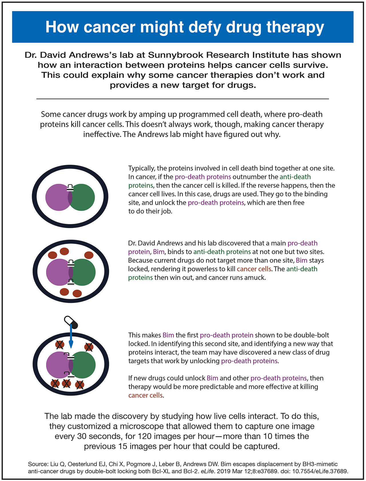
How cancer might defy drug therapy
Dr. David Andrews’s lab at Sunnybrook Research Institute has shown how an interaction between proteins helps cancer cells survive. This could explain why some cancer therapies don’t work and provides a new target for drugs.
Some cancer drugs work by amping up programmed cell death, where pro-death proteins kill cancer cells. This doesn’t always work, though, making cancer therapy ineffective. The Andrews lab might have figured out why.
Typically, the proteins involved in cell death bind together at one site. In cancer, if the pro-death proteins outnumber the anti-death proteins, then the cancer cell is killed. If the reverse happens, then the cancer cell lives. In this case, drugs are used. They go to the binding site, and unlock the pro-death proteins, which are then free to do their job.
Dr. David Andrews and his lab discovered that a main pro-death protein, Bim, binds to anti-death proteins at not one but two sites. Because current drugs do not target more than one site, Bim stays locked, rendering it powerless to kill cancer cells. The anti-death proteins then win out, and cancer runs amuck.
This makes Bim the first pro-death protein shown to be double-bolt locked. In identifying this second site, and identifying a new way that proteins interact, the team may have discovered a new class of drug targets that work by unlocking pro-death proteins.
If new drugs could unlock Bim and other pro-death proteins, then therapy would be more predictable and more effective at killing cancer cells.
The lab made the discovery by studying how live cells interact. To do this, they customized a microscope that allowed them to capture one image every 30 seconds, for 120 images per hour—more than 10 times the previous 15 images per hour that could be captured.
Source: Liu Q, Oesterlund EJ, Chi X, Pogmore J, Leber B, Andrews DW. Bim escapes displacement by BH3-mimetic anti-cancer drugs by double-bolt locking both Bcl-XL and Bcl-2. eLife. 2019 Mar 12;8:e37689. doi: 10.7554/eLife.37689.
This work was funded by the Canadian Institutes of Health Research. The Canada Foundation for Innovation provided infrastructure support for SRI’s Centre for Research in Image-Guided Therapeutics, of which Andrews’ lab is a part.
Original article: Liu Q, Oesterlund EJ, Chi X, Pogmore J, Leber B, Andrews DW. Bim escapes displacement by BH3-mimetic anti-cancer drugs by double-bolt locking both Bcl-XL and Bcl-2. eLife. 2019 Mar 12;8:e37689. doi: 10.7554/eLife.37689.