Nanostructures for biomedical imaging
Naomi Matsuura, Ivan Gorelikov, Kelvin Wan, Siqi Zhu
Nanostructures are a promising new type of contrast agent for clinical medical imaging methods, including magnetic resonance (MR) imaging, X-ray computed tomography (CT), ultrasound and nuclear imaging. Most nanostructures now are simple, single-purpose imaging agents based on spherical constructs. In the next decade, new clinical imaging nanostructures will be designed to be multifunctional platforms that can not only amplify imaging signals at disease sites, but can also be activated by the disease site itself or through means external to the patient to deliver localized therapy.
One goal is to design new types of multimodal nanostructures that can be activated. An example of a potentially activatable multimodal probe developed in our group is an optically tagged perfluorocarbon droplet that can be used as a contrast agent for ultrasound, MR and CT imaging. This multimodal probe may allow us to confirm the labeling of target cells with the droplet on a subcellular level and to trace the fate of the cell in vivo.
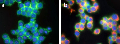
Confirmation of intracellular labeling of macrophage cells with optically tagged perfluorocarbon droplet can be observed with fluorescence microscopy, as shown in the image of fixed unlabeled cells (a) compared to fixed cells labeled with optically tagged perfluorocarbon droplets (b). The intracellular labeling using the perfluorocarbon droplet (as tagged using the orange fluorescent marker) can be seen within the membrane (green) and outside the nucleus (blue).
Customization of nanostructures for biomedical imaging
Our group also designs and customizes novel imaging probes for researchers in biology and medicine. One major bottleneck in the translation of novel nanostructures to useful systems is that the as-synthesized nanostructures must be individually optimized for each application, which usually requires a user-specific surface modification procedure. This is typically difficult and time-consuming, unless a “standardized” surface is used. In our laboratory, we design and develop new coatings for use as versatile platforms by which we can conjugate and modify nanostructures for the end users of the technologies.
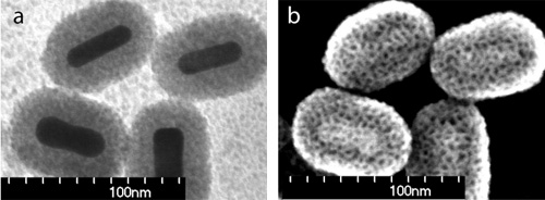
The development of a general and simple method to coat as-synthesized nanostructures will facilitate the rapid adoption, modification and translation of promising new nanostructures into novel and niche applications. One example of a new procedure developed in our laboratory is a robust single-step synthesis method that can be used to coat individual nanostructures with thin highly porous silica shells. The coating provides a functionalizable surface for conjugation to target molecules and the thickness and morphology of the shell can be easily adjusted. This coating method was demonstrated on Au nanorods and CdSe/ZnS quantum dots, with coatings about 15 nm thick with pores about 4 nm in diameter as can be seen in the transmission electron microscope (a) and scanning electron microscope (b) images below.
Reference
- Gorelikov and N. Matsuura, Nano Letters, 8, 369-373 (2008).

