On steady ground
How many of us can say that the work we do is a calling?
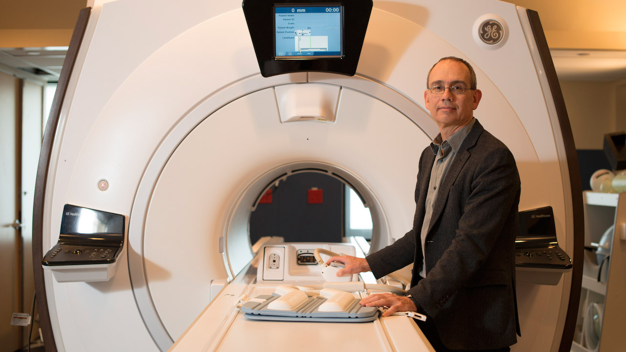
Dr. Graham Wright, a biomedical engineer and director of the Schulich Heart Research Program at Sunnybrook Research Institute (SRI), is one of the lucky ones. He says he was drawn to translational research as a graduate student. “I think the reason I got into it was because I like the hard engineering and physics aspects of medical imaging, with the application of benefitting patients and trying to improve outcomes in health care.”
Wright, who is also a professor in the department of medical biophysics, Institute of Biomaterials and Biomedical Engineering, and Institute of Medical Science at the University of Toronto, has secured a renewal of his Tier 1 Canada Research Chair in Imaging for Cardiovascular Therapeutics. The prestigious award, worth $1.4 million over seven years, recognizes his research leadership and contributions. It entrenches him in his role as head of the cardiovascular MRI research group at SRI. The investment will help him to make advances that have clinical impact, like the technique he developed nearly 30 years ago which uses MRI to characterize blood oxygen content. The method is being applied in patient studies at SickKids and other hospitals to assess cardiovascular disease.
Wright’s lab, which is multidisciplinary, has about two dozen members. The research spans basic biophysics and engineering, preclinical work and clinical studies. The focus is on image guidance for cardiovascular interventions. His team is developing MRI-based tools to guide minimally invasive procedures for the treatment of irregular heartbeats and peripheral arterial disease, in which blood vessels supplying the limbs—most commonly the legs—are blocked.
One of the MRI techniques developed in Wright’s lab has been tested in people with peripheral arterial disease, a condition that afflicts 800,000 Canadians. The study was led by Dr. Trisha Roy, a newly minted PhD co-supervised by Wright and Dr. Andrew Dueck, a clinical researcher at SRI. In it, Roy, who is training to be a vascular surgeon, used MRI to characterize blockages in the leg to predict if they would be difficult to cross during angioplasty, where doctors use catheters and devices like balloons and stents to open clogged vessels. By studying features of the MR signal, they could tell whether an occlusion was likely to be soft, or stiff and difficult to penetrate.
When the participants had their angioplasty procedures their blockages were just as the technique had predicted. Wright notes that the vascular surgeon got through lesions classified as “soft” within a few minutes; those that were deemed “hard” using the technique took much longer to cross—anywhere from 15 to 20 minutes. “In fact, in two of the ‘hard’ lesions they failed to cross, which we expected, and almost all of the ‘hard’ lesions had to be stented. They were more difficult procedures. The MR was quite specific in predicting that,” says Wright.
Being able to show whether an obstruction is pliable or rigid is important in guiding treatment decisions. If it is the latter, then doctors could go straight to bypass surgery, where a piece of a vessel is sewn onto the damaged artery to reroute blood around the blockage, rather than resorting to it after a failed angioplasty attempt. This would result in fewer unnecessary procedures and savings to the health care system.
Wright’s group is also developing a way of using MRI to show the damage to the heart in people with arrhythmia, or abnormal heart rhythm. It is a condition where the heart beats too fast, too slow or irregularly. The aim is to understand better how this damage causes the faulty electrical signalling that is behind irregular heart rhythms, which can lead to sudden cardiac death.
Looking ahead, Wright says a main goal is to bring discoveries made in the lab closer to clinical practice. “Our single biggest push will be to get some of these tools into larger patient studies, which will hopefully lead to broader clinical adoption,” he says.
This year has been stellar for Wright, who in the spring was elected a Senior Fellow of the International Society for Magnetic Resonance in Medicine. But for all the accolades on his CV, the accomplishment of which he is most proud is the success of his trainees—past and present. “It’s very exciting to see them go out from the lab and become established in their careers. Almost all of them have stayed in the general area of medical research, technology development or medicine, with a stronger understanding of the underlying challenges on the technical side. That’s the single biggest thing that I can look back on and say has been rewarding.”
In a nutshell
- Dr. Graham Wright secured a renewal of his Tier 1 Canada Research Chair in Imaging for Cardiovascular Therapeutics.
- The award supports his research into using MRI to guide cardiovascular interventions.
- The aim is to move discoveries made in the lab closer to clinical practice.

