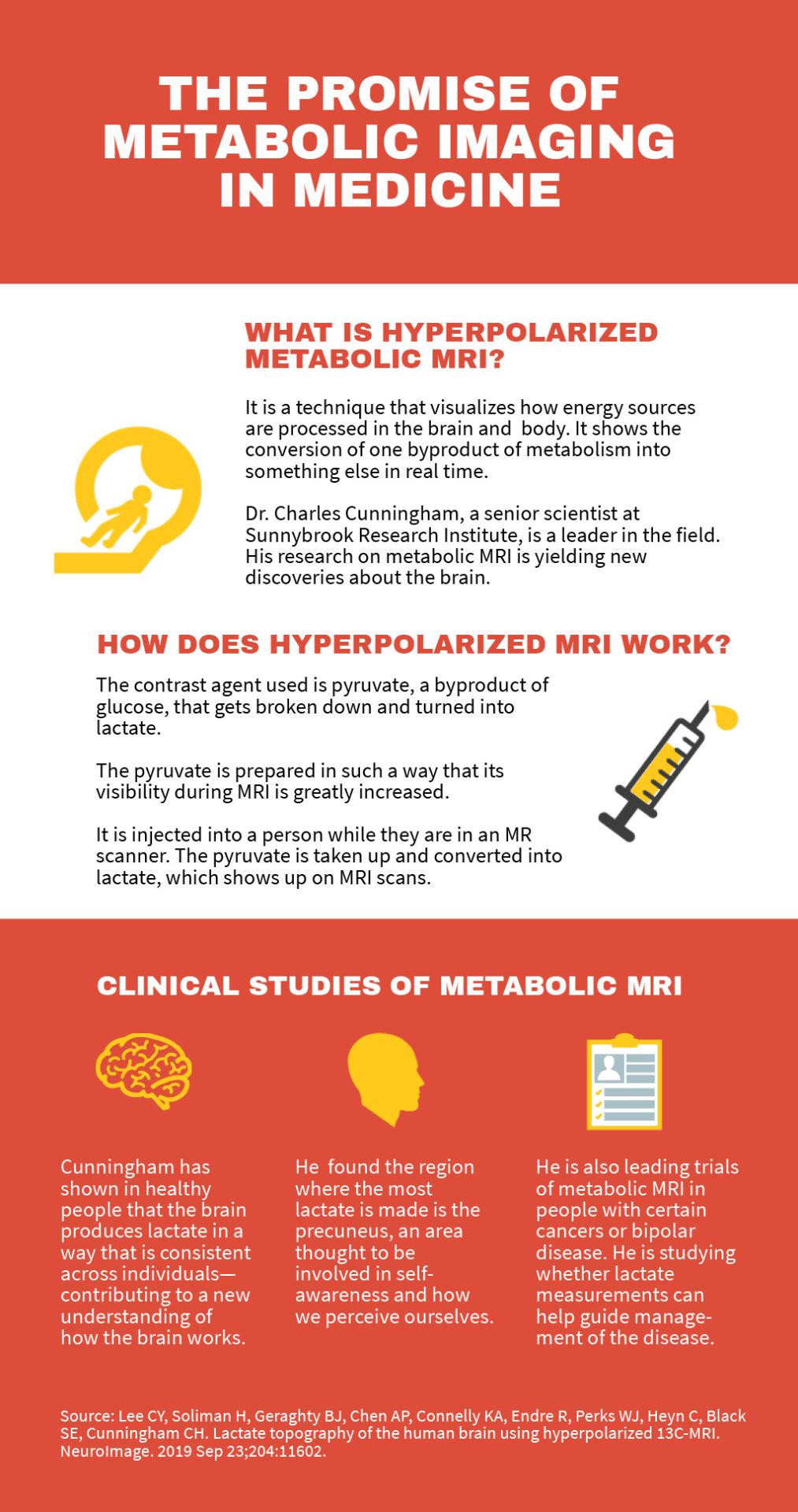
The promise of metabolic imaging in medicine
One of the most energy-hungry organs, the brain uses about 25% of the body’s total caloric requirements—this despite weighing only 2% of its mass. Understanding brain energy metabolism is leading to new discoveries into what goes on in our grey matter. These findings, like one made by Sunnybrook Research Institute (SRI) senior scientist Dr. Charles Cunningham, are upending long-held views about substances made in the brain as the body breaks down food. He published a study showing that people make a product of metabolism called lactate in their brains, and that they make it in specific regions.
“The most exciting thing we’ve found is that the brain does produce lactate inside the brain tissue. It has a distinct spatial pattern, and that pattern is really consistent across people, which makes me think it’s tightly controlled in the brain. It’s like a ‘map’ of metabolism in the brain that’s potentially a totally new way of looking at how the brain works,” says Cunningham of the study, published on Sept. 23, 2019, in the journal NeuroImage.
It was believed that the brain only produces lactate, which is derived from blood sugar or glucose, when oxygen is insufficient. Under normal conditions, the brain has excellent blood flow and therefore plenty of oxygen, so there should be very little lactate in the brain, or so the thinking goes. The notion that the brain creates and uses lactate is somewhat controversial; Cunningham’s discovery adds to the evidence supporting this idea.
He is a leader in metabolic MRI, also called hyperpolarized MRI, a technique that enables chemical reactions in tissue to be seen noninvasively in real time. It shows what no other imaging tool can show: the conversion of one byproduct of metabolism into something else in the body or brain as it happens.
The contrast agent used in this technique is pyruvate, a byproduct of glucose that gets broken down and turned into lactate. The pyruvate is prepared in a strong magnetic field so that its visibility during MRI is greatly increased. It is injected into a person while they are in an MR scanner. Then, the pyruvate is taken up and converted into lactate. All of this shows up on MRI scans.
Cunningham and colleagues found that the area where the most lactate is made is the precuneus [pre-kyoo-nee-us], which is somewhat buried in the middle of the brain. Relatively little is known about the precuneus, but it’s believed to be involved in self-awareness and how we perceive ourselves.
“When you instruct people to lay there, close their eyes and not think about anything, the precuneus is the most metabolically active area in the brain,” says Cunningham. “It’s thought that when your brain is not doing anything else—you’re conscious and self aware, and that’s the only thing you’re doing—the precuneus is one of the main brain regions involved in that. It seems to be the part of the brain that’s creating the feeling of being who you are. These images really may heighten the research effort into understanding what the precuneus is doing.”
Altered metabolism: a window into disease
The ability to measure lactate in tissue has important implications for treating various conditions. Cancer cells consume about 15 times more glucose than do normal cells; production of lactate is the major reason for this. “Parts of tumours tend to produce very high lactate, because lactate has several advantages for tumour cells to grow. It’s both a signalling molecule and a way for cells that don’t have oxygen to consume energy without depleting the glucose that’s needed for the other cells to survive. It turns out that high lactate in a tumour can be used to predict the outcome of a [cancer] patient,” says Cunningham, who is also an associate professor of medical biophysics at the University of Toronto.
By showing how much lactate there is in a tumour, for example, metabolic MRI could be used to predict how difficult a cancer will be to treat. It could also help doctors determine right away whether a tumour is responding to treatment.
Along with Dr. Hany Soliman, a cancer researcher at SRI and radiation oncologist at Sunnybrook, Cunningham is testing this technique in several clinical trials, including one of people with metastatic brain cancer, where cancer has spread from its original site to the brain.
“It’s a bad sign if the tumour has high lactate. We could measure lactate being produced in brain metastases, and it would tell us before patients were treated how likely they are to respond to treatment,” says Cunningham.
He explains that a lot of lactate translates into a greater chance of poor treatment response. “Also, if the lactate changes after treatment, then it would be a marker for response to therapy,” says Cunningham.
Soliman notes there are no indicators for early response to therapy that can help doctors to determine whether someone with metastatic brain cancer is on the right course. It can take weeks or months to see if a tumour shrinks in size, to thus conclude that a treatment is working. “There is no other marker, only anatomical volumetric changes,” he says.
Cunningham is also leading clinical studies evaluating metabolic MRI in people with cancers of the prostate and breast, and locally advanced cervical cancer. Like with the brain cancer trial, he is studying if using metabolic MRI to measure lactate in tumours can predict aggressive disease and show early treatment response.
He was the first to show that hyperpolarized MRI could be used to visualize biochemical reactions in normal human hearts. He is now studying people with enlarged hearts. This condition increases the risk of heart failure, where the heart is too weak to meet the body’s pumping needs. In this clinical trial, the first of its kind, he aims to learn whether pyruvate metabolism can be used to predict disease progression in patients at risk of heart failure so that treatment can be personalized. For instance, people who are at increased risk of heart failure can be prescribed certain drugs to improve heart function. The trial follows Cunningham’s pioneering work in figuring out just how to use hyperpolarized MRI in the heart, a challenging feat owing to the muscle’s motion and fast blood flow.
Another area in which metabolic imaging could prove useful is the study and management of bipolar disorder. A mental illness resulting in extreme mood swings that include emotional highs and lows, bipolar disorder can be debilitating. About 1% of Canadians will experience it in their lifetime.
Cunningham is working with Dr. Benjamin Goldstein, a senior scientist in the Hurvitz Brain Sciences Research Program at SRI and a psychiatrist at Sunnybrook, to study whether measuring lactate can be used to assess response to therapy in youth with bipolar disorder.
Goldstein notes that problems with energy metabolism and dysfunction of the mitochondria, which are like energy factories inside the cells, are thought to be the main contributors to bipolar disorder. Moreover, research shows that brain lactate is elevated in people with the disease. “This technique would therefore provide unique insights regarding lactate in the brains of teens with bipolar disorder,” says Goldstein of the clinical trial on which they are collaborating.
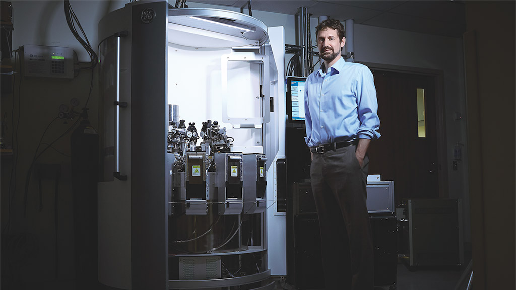
To date, only nine other sites in the world have done human studies of hyperpolarized carbon-13 MRI. Sunnybrook Research Institute is the only institution in Canada with a SPINlab polarizer, the equipment on which clinical studies of this imaging technique are performed. It also has a research hyperpolarizer. Both are part of SRI’s Centre for Research in Image-Guided Therapeutics.
Cunningham has spent more than a decade developing the hardware and image acquisition methods for metabolic MRI, always with the view to translating the research clinically. He says with a smile, “It actually seems to be showing a lot of promise for being able to guide treatment in patients. That was always the plan. That’s what I was shooting for.”
Read text-only version of above infographic
The promise of metabolic imaging in medicine
What is hyperpolarized metabolic MRI?
It is a technique that visualizes how energy sources are processed in the brain and body. It shows the conversion of one byproduct of metabolism into something else in real time.
Dr. Charles Cunningham, a senior scientist at Sunnybrook Research Institute, is a leader in the field. His research on metabolic MRI is yielding new discoveries about the brain.
How does hyperpolarized MRI work?
The contrast agent used is pyruvate, a byproduct of glucose, that gets broken down and turned into lactate.
The pyruvate is prepared in such a way that its visibility during MRI is greatly increased.
It is injected into a person while they are in an MR scanner. The pyruvate is taken up and converted into lactate, which shows up on MRI scans.
Clinical studies of metabolic MRI
Cunningham has shown in healthy people that the brain produces lactate in a way that is consistent across individuals—contributing to a new understanding of how the brain works.
He found the region where the most lactate is made is the precuneus, an area thought to be involved in selfawareness and how we perceive ourselves.
He is also leading trials of metabolic MRI in people with certain cancers or bipolar disease. He is studying whether lactate measurements can help guide management of the disease.
Source: Lee CY, Soliman H, Geraghty BJ, Chen AP, Connelly KA, Endre R, Perks WJ, Heyn C, Black SE, Cunningham CH. Lactate topography of the human brain using hyperpolarized 13C-MRI. NeuroImage. 2019 Sep 23;204:11602.
Cunningham’s research was supported by Brain Canada, the Canada Foundation for Innovation, Canadian Cancer Society, Canadian Institutes of Health Research, and the Ontario Ministry of Economic Development, Job Creation and Trade.
In a nutshell
- Dr. Charles Cunningham has shown in healthy people that the brain produces lactate in a way that is consistent across individuals—contributing to a new understanding of how the brain works
- He is leading clinical trials of metabolic MRI in people with enlarged hearts, certain cancers and bipolar disorder.
- In people with cancer or bipolar disorder, he and his colleagues are studying whether using the technique to measure lactate, a byproduct of metabolism, can help assess response to therapy and guide management of the disease.

