Big-Picture Physicist
Specialist’s breadth of knowledge brings creative solutions to research lab
By Dilys Chan
What is digital mammography?
Now used all over the world, digital mammography takes and stores electronic pictures of the breast. The digital images can be viewed conveniently on a monitor, e-mailed and adjusted for brighter, sharper image. When compared with standard film mammography, research shows that it reduces wait times for imaging, makes long-distance consultations easier and helps detect cancer earlier in certain groups of women.
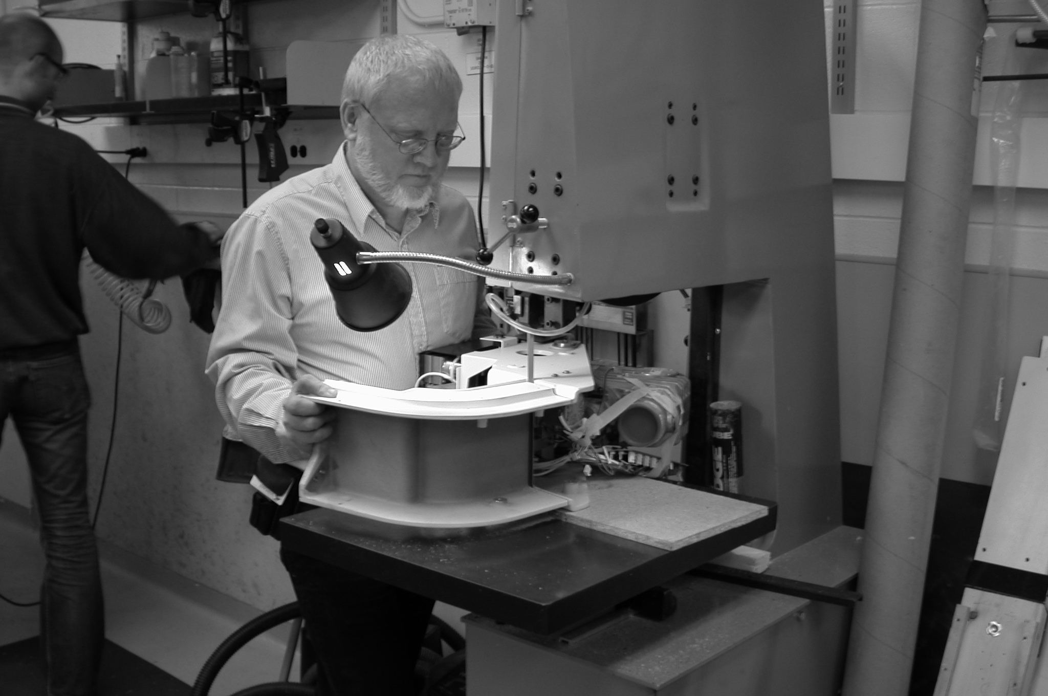
No two days are the same for Gord Mawdsley, a medical physicist in the lab of senior scientist Dr. Martin Yaffe, whose research group studies the detection and nature of breast cancer. After more than 15 years of working at Sunnybrook Research Institute (SRI), he knows a little bit about everything that goes on in Yaffe’s lab.
Mawdsley studied physics, receiving a bachelor of science from the University of Toronto. He also took three years of machining and electronics in high school. His job, as he puts it, involves the “practical application of physics.”
For example, to study the distribution of cancer cells and biomarkers in tissue, the group is developing pathology techniques in which a whole specimen of tissue removed during surgery is imaged. In normal pathology, the tissue is sampled, and small sections are put on a one-by-three-inch slide.
In Yaffe’s lab, however, they slice, process and mount the entire breast tissue samples onto slides measuring up to five by seven inches. Mawdsley designed and built a microscope that could handle the large slides and image over 50 gigabytes of data per slide. He also developed a small computed tomography scanner so that the tissue could be imaged before it was sliced, enabling the alignment of each histology image with X-ray images. A problem that arose was that every piece of tissue mounted had a different angle to it, making it difficult to line up the slides for examination and comparison.
The solution, identified by another group at SRI, involved black spaghetti.
A Novel Solution
“It’s real spaghetti; you can eat it, and it’s been dyed with cuttlefish ink, which has a lot of iron in it so you can see it in an MR [magnetic resonance] scanner.” While the spaghetti showed up well in images, pushing it through tissue caused it to bend. Mawdsley’s idea was to push spaghetti through the centre of biopsy needles, typically used to remove small amounts of breast tissue. After pulling the needles out of the sample, the black spaghetti is left behind in the tissue.
The result? “Nice and straight,” he says.
Other parts of Mawdsley’s job involve keeping the lab group organized, communicating the lab’s needs with architects who are planning new facilities, meeting purchasing guidelines for equipment, working with clinicians to set up clinical trials and training students.
A Multifaceted Role
In addition to his work at SRI, Mawdsley acts as a mammographic physics consultant for the Ontario Breast Screening Program, ensuring that image quality and radiation dosage are appropriate.
“It keeps us out of the ivory tower,” he says. “That’s very useful, because a lot of the physics measurements we do are optimized in the lab, and then in the field, we find out that there are bigger problems that an understanding of basic physics helps solve.”
Mawdsley’s work also exposes him to SRI’s commercialization activities and partnerships with the private sector. Currently, the research group collaborates with GE Healthcare to evaluate their developments in producing 3-D X-ray images of the breast. He’s also participated in some long-term achievements, notably in taking digital mammography detectors from prototype to commercialization. (See sidebar.) Yaffe’s group was involved in the early development of digital mammography and is now playing an instrumental role in developing international quality standards.
“It’s kind of exciting to have built a prototype machine 15 years ago, and then have a company use our designs to make a digital mammography product,” he says. “And now, to see it replacing film mammography.”
Infrastructure for the research into digital mammography and breast pathology was enabled by investment from the Canada Foundation for Innovation and the Ministry of Research and Innovation.