Field of view
By Alisa Kim
On Jan. 10, 2014, more than 100 faculty, trainees and staff from Sunnybrook Research Institute (SRI) gathered to attend the third annual Centre for Research in Image-Guided Therapeutics (CeRIGT) Research Day.
The event showcased the science of CeRIGT, a $160-million centre that was established by federal funding through the Canada Foundation for Innovation and cemented by provincial and private sector investment. Here, researchers are exploiting SRI's strength in imaging and biology to invent new and better ways to detect, diagnose and treat complex conditions including cancer, cardiovascular disease, musculoskeletal disorders and dementia.
Dr. Michael Julius, vice-president of research at SRI and Sunnybrook, welcomed everyone and highlighted the mandate of CeRIGT researchers: translating collaborative, multidisciplinary research into clinical impact. "CeRIGT is dedicated to convergence, to breaking down silos so that physicists, biologists, engineers and clinicians work together. Our people are our most precious asset. The centre will accelerate successes we've shepherded to this point and many more to come," said Julius.
Dr. Frank Prato, who is the assistant scientific director of the Lawson Health Research Institute in London, Ontario, gave the keynote lecture on strengths and potential applications of fully integrated positron emission tomography (PET) and magnetic resonance imaging (MRI).
He noted the complementarity of the modalities: MRI produces detailed structural images while PET does molecular imaging, which gives functional or physiological information such as metabolism or microscopic pathologies. Moreover, taking images from each modality simultaneously addresses the problem of patient motion when images are obtained from different machines. The fusion technology expedites imaging procedures and improves diagnostic accuracy, said Prato. Research applications include development of PET probes to monitor a cancer patient's response to therapy; and, in cardiology, detecting inflammation implicated in heart failure and vulnerable plaques, the culprit behind heart attacks.
Dr. David Andrews, SRI's director of Biological Sciences, described how he is using an automated microscope that he helped develop with industry to study protein-protein interactions in live cells. The instrument makes high-resolution images—up to 100,000 a day, by itself—which allow him to identify sites of potential therapeutic interaction between the proteins that regulate cell death, and validate them as targets. Andrews uses the technology to test small molecules; it is of interest to drug companies because it not only shows how a drug works, it also is a fast and inexpensive way to rule out drugs that don't work.
Dr. Cari Whyne, director of the Holland Musculoskeletal Research Program, outlined how she is using finite element modeling, a computational bone-modeling technique, to visualize stresses and strains to damaged and undamaged areas. She noted that understanding load thresholds could help reduce the risk of fracture in people with cancer that has spread to bone. Finite element modeling also provides 3-D representations of strains placed throughout the skull; understanding the flow of mechanical loads could improve reconstruction methods for patients with craniofacial trauma, she said. Whyne also spoke about promising preclinical research on the use of lithium to improve healing, which is delayed or impaired in 10% of patients with fractures.
In all, there were 12 talks covering the full spectrum of translational research: from basic science, to preclinical testing and human trials, to evaluation of outcomes.
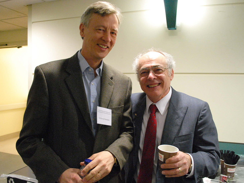
Dr. Kullervo Hynynen, director of Physical Sciences and project lead of CeRIGT, closed the day by thanking all for their participation and remarked that the talks sparked several ideas for collaboration within his research program.
For more about CeRIGT, visit sunnybrook.ca/research/cerigt


