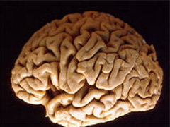
Head Injuries Result In Widespread Brain Tissue Loss One Year Later
“This is an important finding as TBI is one of the most common forms of disability,” said Dr. Brian Levine, Senior Scientist at Baycrest’s Rotman Research Institute and lead author of the study which is published in the March 4, 2008 issue of Neurology, the medical journal of the American Academy of Neurology.
TBI causes both localized damage through bruises or bleeds, as well as more diffuse damage through disconnection of brain cells, which ultimately causes cell death. The localized damage is easier to detect with the naked eye than diffuse damage. Yet both kinds of damage contribute to difficulties with concentration, working memory, organizing and planning (vital skills for holding a job), and mood changes often experienced by people following TBI.
According to Dr. Levine, “It can be hard to determine why patients are so disabled, and this study offers a clue to the nature of the brain damage causing this disability.”
In the study, 69 TBI patients were recruited from Sunnybrook Health Sciences Centre, Canada’s largest trauma centre, one year after injury. Eighty percent of the patients sustained their injury from a motor vehicle accident. Injury severity was determined by the depth of coma or consciousness alteration at the time of the initial hospitalization. Some patients had minor injuries and were discharged immediately, whereas others had more severe injuries with extended loss of consciousness lasting weeks. Twelve healthy, non-injured participants were recruited as the comparison group.
Subjects’ brains were scanned with high resolution magnetic resonance imaging (MRI) which provides the most sensitive picture of volume changes in the brain. In addition to using an expert radiologist’s qualitative reading of the MRI scans, which is the standard approach used in hospitals and clinics, the researchers processed the images with a computer program that quantified volumes in 38 brain regions.
The computerized analysis revealed widespread brain tissue loss that was closely related to the severity of the TBI sustained one year earlier. “We were surprised at the extent of volume loss, which encompassed both frontal and posterior brain regions,” said Dr. Levine. Brain tissue loss was greatest in the white matter (containing axons which can be compared to telephone wire interconnectivity), but also involved grey matter (containing the cell bodies important for information processing).
Investigators were surprised to find that volume loss was widespread even in TBI patients who had no obvious lesions on their MRI scans. Even the mild TBI group contributed to the pattern of volumetric changes such that this group was reliably differentiated from the non-injured, healthy group.
“A significant blow to the head causing loss of consciousness can cause extensive reduction of brain tissue volume that may evade detection by traditional qualitative radiological examination,” Dr. Levine noted.
He is leading follow-up studies on the same group of TBI patients to examine more closely the significance of localized white and grey matter volume loss on behaviour.
The research team for the Neurology paper included Dr. Sandra Black, Neurosciences Program Research Director at Sunnybrook Health Sciences Centre, Brill Professor of Neurology, Department of Medicine, at Sunnybrook and University of Toronto, and Senior Scientist at Sunnybrook and the Rotman Institute at Baycrest. Both Baycrest and Sunnybrook are fully affiliated with the University of Toronto.
The study was supported by grants from the Canadian Institutes of Health Research and the National Institutes of Health.
PDF / View full media release »


