


Imaging Student Wins Prize for Novel Catheter
By Jim Oldfield
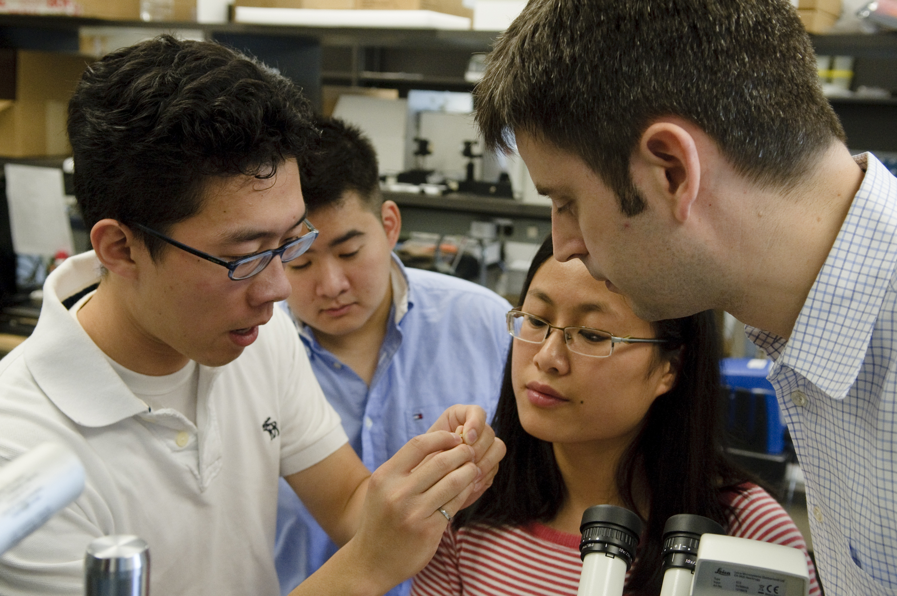
Brian Li placed first in the Imaging for Cardiovascular Therapeutics poster competition at the 2010 Imaging Network Ontario research symposium, held at the University of Toronto on February 2. Judges selected four first-place posters, one from each segment of the symposium, from a province-wide pool of 150.
"I was very surprised and humbled to be in a category with those receiving similar honours," said Li, a first-year U of T medical student in the lab of Sunnybrook Health Sciences Centre resident physician and researcher Dr. Brian Courtney.
Li's project is a hybrid intravascular ultrasound/optical coherence tomography catheter for imaging vascular disease. The combined catheter provides detailed structural and compositional information on vulnerable plaque—a type of lipid accumulation inside an artery wall that is hidden under thin-plaque caps. Spontaneous rupturing of these plaque produce blood clots, and they are the leading cause of heart attacks. Currently undetectable in patients, these plaque can strike without warning.
"Intravascular ultrasound alone is one method to image plaque, but while it offers good depth and penetration, its resolution and ability to distinguish which plaque are vulnerable is limited," said Courtney. "So we've been trying to combine it with other modalities, such as optical coherence tomography, which has limited penetration depth but great resolution and contrast."
To date, only one other research group in the world has developed a system that compactly combines the two technologies in one catheter, but at 2.4 millimetres in diameter it is still too large to be practical for intravascular imaging. Li and Courtney's catheter, in contrast, is just 1.3 millimetres across, allowing access to most coronary arteries. Their lab has tested the device in diseased vessels from human cadavers; the next step is in vivo imaging in animal models of cardiovascular disease.
Courtney and Li's design co-aligns the device's ultrasound and optics, enabling complementary imaging of the exact same place in the vessel. This is a huge improvement on the strategy applied by previous researchers, whereby they imaged first with an ultrasound catheter followed by a second separate catheter for optical coherence tomography—an approach that increases procedure time and patient risk through multiple insertions. Repeated catheterizations can also alter the plaque site between image acquisitions and create uncertainty regarding plaque structure and which area is being imaged by each technique.
Li said there is work to be done in testing the device at the preclinical stage, but he believes it has great potential to aid diagnosis and treatment planning in patients. As such, the technology merges not only imaging modalities, but Li's two main interests: "It's not just about developing new technology—it's working toward clinical impact," said Li. "In that respect, working with Dr. Courtney on this device has been perfect."
Courtney and Li's work was funded by Canadian Institutes of Health Research, Canada Foundation for Innovation, Health Technology Exchange, BioDiscovery Toronto and the Ministry of Research and Innovation.