Ringing True
By Laura Pratt
Sometimes, science sings.
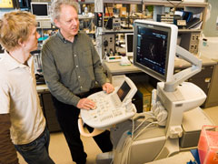
Dr. Peter Burns, a senior imaging scientist at Sunnybrook Research Institute, has proved this definitively with research that concludes that microbubble-enhanced ultrasound-his innovation-is at least equal to computerized tomography (CT) and magnetic resonance (MR) scanning in its ability to qualify the nature of a lesion in the liver. A paper published in the January 2007 edition of Radiology, images from which made the journals cover, compares Burns's clinical trial results of CT, MR and microbubble-enhanced ultrasound scanning of 135 patients. It confirms his assumptions that his method-already celebrated for being safer, more prevalent and more economical-is every bit as technically proficient as those previously believed to be the only option for patients whose chronic liver disease necessitates regular monitoring to check for the development of tumours that may prove life threatening.
People with chronic liver disease, such as that arising from such common conditions as hepatitis infection, require continuous surveillance. Depending on the case, this might mean being scanned once every six months. Often the patients are young people, for whom the prospect of large doses of radiation-inherent in CT scans-is dire, and there may not be the resources for MR scans, which are less widely available and prohibitively expensive. This is particularly critical news in Toronto, where the large Asian community means a huge segment of the population is diagnosed with Hepatitis B (endemic in some Asian countries).
That leaves ultrasound, the undisputed leader on the imaging landscape. (The number of exams conducted with ultrasound exceeds all other scanning methods for diagnosis combined.) Historically, ultrasound has proven the best surveillance modality for looking at liver disease-to a point. This modality can find masses only in the liver (and between 10% and 20% of us have them, frequently harmless); they can't identify their nature. As such, ultrasound was never a viable option for monitoring liver disease, because it lacked the contrast agents whose revelation of the pattern of blood flow inside a lesion makes its complete diagnosis possible. Ultrasound without contrast is basically relegated to the role of seeing, but not knowing.
Until recently.
Working with a pharmaceutical firm from Germany and a high-tech ultrasound imaging company from the United States, Burns's ultrasound imaging research lab developed a method by which a contrast agent could be used-for the first time ever-for ultrasound, a sea change that resulted in an entirely new imaging modality in medicine. That imaging method, harmonic and pulse-inversion imaging, was licensed to the ultrasound industry by Sunnybrook in 1999. It's now widely available on ultrasound scanners worldwide.
Here, a cloud of microscopic bubbles, made using high-technology pharmacological fabrication techniques that produce tiny bubbles (three to five millionths of a metre across-about the same size as a red blood cell) of gas coated with lipids, is injected into the blood stream where they circulate, harmlessly. The bubbles ring like bells in response to sound, and produce overtones or harmonics. A specially developed ultrasound scanner detects only the harmonics of the echoes. The technology is so sensitive it can trace the pattern of individual bubbles 10 cm into the liver, in real time.
This revolutionary new method for diagnosing the nature of abnormalities in patients suffering from liver disease-a condition Health Canada acknowledges in March with its assignment of Help Fight Liver Disease Month-was approved by Health Canada in 2003. It has been music to the ears of the medical science community worldwide.
Burns' research was developed with support from the Canadian Institutes of Health Research and the Terry Fox Foundation of the National Cancer Institute of Canada.
PDF / View full media release »


