
Fine-tuning imaging to enhance craniofacial implants for patients
Reconstructing the craniofacial skeleton after trauma often requires implants that must match the facial structure of the patient to restore both function and cosmetic appearance. Holland MSK researchers are working to improve imaging visualization of thin bones in the face, in order to enhance the design of these implants.
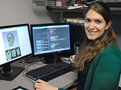
Cari Whyne, senior scientist, Michael Hardisty, research engineer, Sunnybrook Research Institute, and Amani Ibrahim, a student working in the orthopaedic biomechanics laboratory, are developing software to improve the image-processing technology. They are collaborating with Calavera Surgical Design, a startup company out of Sunnybrook Research Institute, which has developed a process that uses patient-specific molds from CT (computed tomography) scans to shape mesh into customized craniofacial implants.

Clinical CT scans provide only limited visualization of finer structures such as the orbital floor of the eye socket or bones near the nasal cavity. The researchers whose work is funded by the Ontario Centres of Excellence and FedDev Ontario, are using filters from point data to sharpen and de-blur these images to create smooth continuous surfaces which can be 3D printed into patient specific molds.
At this year’s 14th Annual Imaging Network Ontario Symposium, Amani Ibrahim received the first prize “Summa cum Laude” award in the bone and joint category for her oral presentation, “Image Processing Software for Designing Custom Craniofacial Implants.”