
Behind the research: Developing artificial intelligence applications to speed up cancer diagnosis
Sunnybrook research teams are developing advanced machine learning algorithms to automatically localize cancerous tissue in patient samples and ultimately speed up pathology turnaround times.
The work was recently recognized internationally with two students from senior scientist Dr. Anne Martel’s lab placing second in two arms of the SemiCOL Challenge, a global AI digital pathology challenge.
Colorectal cancer is the fourth most commonly diagnosed cancer in Canada, and was the second leading cause of death from cancer in men and the third in women in 2022.
“In order to diagnose a patient with colorectal cancer, an expert pathologist analyzes surgically removed tissue samples that are magnified and scanned,” says Tony Xu, a PhD student at SRI in the Martel lab and recent SemiCOL awardee. “The scanned images are difficult to analyze because of their immense size (often over 100,000 x 100,000 pixels).”
To alleviate this and assist pathologists in finding cancerous tissues in the images, the semi-supervised learning for colorectal cancer detection, or SemiCOL challenge is an international online competition about using machine learning (ML) methods to automatically localize tissues that are associated with colorectal cancer.
The researchers explain that the challenge to developing successful ML algorithms is training the large data sets.
“While a large amount of raw data is often available in medical settings, it is both expensive and time-consuming for medical experts to create detailed annotations for this data,” says Matthew McNeil, also a PhD student in the Martel lab and recent SemiCOL awardee. “The scarcity of labels created by medical experts is one of the key issues in applying ML algorithms to medical data.”
Two recently popular branches of ML called “self-supervised learning” and “semi-supervised learning” aim to train algorithms with little to no labels. These methods operate by teaching ML models to recognize structures, patterns and other common image qualities from unlabeled data. Once they are trained on such data, these models can then be applied to detection tasks, such as the one in the SemiCOL challenge.
Competition was fierce, with both academic research labs and companies already working in the area of GI pathology taking part. Despite this, Xu and McNeil were able to use cutting edge AI algorithms and their existing expertise in digital pathology to achieve second place in the challenge, securing 2000Euro in prize money. They also presented their results at the 19th European Congress on Digital Pathology.
“With funding provided by an INOVAIT Pilot Fund, we have amassed a large database of colonoscopy images here at Sunnybrook,” says Dr. Martel. “In collaboration with Dr. Kenneth Craddock and Dr. Lina Chen, experts in GI pathology, we will use this data to train an AI model capable of locating cancer, allowing pathologists to work more accurately and efficiently. The models trained for the Semicol challenge provide an excellent springboard for this.”
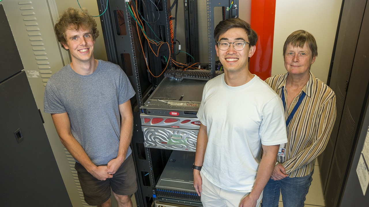
The work was made possible thanks to the Artificial Intelligence Platform for Precision Medicine (AIPPM) at SRI supported by a recent Canada Foundation for Innovation (CFI) grant. The heavy-duty AIPPM servers enabled the team to accelerate experiments and move past memory constraints to explore new deep learning architectures.
Dr. Anne Martel is hoping that the AI models developed for colonoscopy, and other applications such as the detection of cancer in breast and lymph node tissue biopsies, will soon be translated into the clinic. “These automated algorithms have the potential to decrease patient turnaround and speed up pathologist workflows, opening up their time for other essential tasks”.