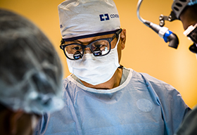
How the Breast Rapid Diagnostic Unit works
The clinic provides rapid assessment and diagnosis for individuals with an abnormality on a mammogram, breast ultrasound or a clinical finding that is highly suspicious of breast cancer.
The Breast Rapid Diagnostic Unit (RDU) also features a dedicated nurse navigator who guides and supports each individual through the assessment process.
Individuals also benefit from the combined expertise of the Odette Cancer Centre breast care team and other breast-dedicated specialists in the departments of medical imaging and anatomic pathology at Sunnybrook.
Physician referrals are required.
Who is eligible for the RDU?
Before Your Breast Imaging Appointment
A Nurse navigator will contact you to arrange the following:
- If your initial imaging is not from Sunnybrook or it is not available on a local or on the regional imaging database, you will be asked to contact the place where your imaging was done to pick up a CD that has your imaging on it.
- Set up an appointment for further imaging and biopsy.
- Provide instructions for preparation for the appointment.
Prior imaging review:
- Before your RDU appointment, a radiologist at Sunnybrook will review the imaging to ensure the findings meet the eligibility criteria. If the imaging findings are not concerning for malignancy (cancer), you may be seen outside of the RDU, as the RDU appointments are reserved for findings that are suspicious for malignancy and likely to require biopsy.
- We compare your recent breast imaging with other imaging that you had in previous years. We use the comparison to help decide who needs additional work-up and biopsy.
Preparing for your appointment:
- Tell the nurse navigator about any blood thinners you take (eg. Advil, Aspirin, Naproxen, Warfarin, Plavix or Apixaban) and if you have any allergies to local anesthetic or latex.
- You are allowed to eat before your appointment. A healthy breakfast is encouraged to help prevent you from feeling lightheaded during the procedures.
- Please contact the RDU nurse with any concerns
Please contact RDU nurse if there are any concerns or if you are unwilling to undergo biopsy. The RDU appointments are designed specifically to provide faster testing and diagnosis for patients who need a biopsy. If you do not need a biopsy, the nurse will let you know and the breast imaging department will phone you to come in for further imaging only.
Your Diagnostic Imaging and Biopsy Appointment
Here is the outline for most imaging and biopsy appointments:
- Check in on M6 Breast Imaging Reception (the first reception area).
- The Nurse navigator will meet with you to take a clinical history and review what will happen during the appointment.
- You will change into a gown and the technologist will do a mammogram. The radiologist will check the mammogram and may ask for additional specific mammographic views to further assess the breast tissue.
- As you may have experienced previously, mammograms require compression in order to spread out the breast tissue as best as possible such that an accurate diagnosis can be made. For this reason, mammograms may be uncomfortable but technologists do their best to minimize this discomfort and address your concerns.
- Once the mammogram and possible views are completed, the images will be reviewed and then you will be taken for an ultrasound by a technologist. This is done while you are lying down on a stretcher.
- The technologist will review the imaging with the radiologist and the radiologist will then come and speak to you and will likely also do another scan themselves.
- Decision for biopsy will be made and the radiologist will perform the biopsy with the assistance from the technologist.
- Most of the biopsies are done under ultrasound guidance but some are done using mammographic guidance. This will be discussed with you prior to the procedure.
- Sometimes enlarged lymph nodes are seen in the axilla and in addition to a breast biopsy, a fine needle biopsy of the lymph node may be performed.
- After the biopsy, you will be given post-biopsy instructions and an appointment to return for results within 24-72 hrs.
- Sunnybrook is a teaching hospital. There may be a trainee physician assisting, scanning or performing part or all of the procedure under close supervision by the radiologist.
Frequently Asked Questions
What is an ultrasound-guided biopsy?
- An ultrasound-guided biopsy is a procedure performed under local anesthetic (numbing to the area). Ultrasound is used to guide a biopsy needle to get small pieces of tissue from a mass (abnormal cells). The tissue is sent to the lab to be assessed under a microscope and for other specialized tests to determine a diagnosis.
- You will be lying down on the same stretcher on which the ultrasound was done, in the same position (arm up).
- The radiologist will mark the site for biopsy and then clean the skin with sterile solution and place sterile drapes around the sterilized area.
- Local anesthetic will be injected at the site with a small needle. 1% Lidocaine is typically used.
- A very small skin incision (cut) will be made and a biopsy needle will be put into the site in question. The doctor will guide the needle and use the needle to take a small core of tissue. The needle will make a loud snapping noise.
- The needle will be taken out. The tissue will be removed from the needle.
- The needle will be used to take further samples from the breast, typically 4-5 total.
- A metal clip may be placed at the biopsy site to help the radiologist find the area again if needed.
- The area will be cleaned and dressed by the technologist.
- If a metal marker clip is placed, you will have a post-procedure gentle mammogram for mammographic documentation.
What is a stereotactic biopsy?
- An stereotactic biopsy is a procedure done under local anesthetic (numbing) to get small pieces of tissue from a calcification using a biopsy needle and mammographic guidance to direct the needle. The tissue is sent to the lab to be looked at under a microscope and for other specialized tests to determine a diagnosis. You will be either lying down on your stomach or sitting up in a chair in a room with a stereotactic biopsy machine.
- The breast will be compressed and the technologist will get initial localizing mammographic views.
- The radiologist will target the biopsy site on the images and send the coordinates to the biopsy device.
- The radiologists will clean the skin and local anesthetic will be injected at the site with a small needle. 1% Lidocaine is used for skin and superficial tissue and a deeper injection of 1% Lidocaine with epinephrine is used to help control any bleeding. Further localizing images may be obtained.
- A very small skin incision (cut) will be made and the doctors will advance the biopsy needle to the site in question in your breast. Images may be taken at this time.
- When positioning is confirmed, the biopsy samples will be taken, most commonly using a vacuum-assisted biopsy device. This device will rotate on its axis to take multiple samples of the area around the “clock”.
- The biopsy needle will be removed.
- The samples will be removed from the biopsy device and imaged to ensure the calcifications in question have been adequately sampled. It is possible that further samples will be required following this imaging.
- A metal marker clip may be placed at the biopsy site to help the radiologist find the area again if needed.
- The area will be cleaned and dressed by the technologist.
- If a marker clip is placed, a post-procedure gentle mammogram will be obtained for mammographic documentation.
Why do we use marker clips?
A marker clip a metal clip made of titanium that is left in the breast to help the radiologist find the area again in the breast if needed for surgery or monitoring.
- The marker clip ensures that we know exactly which area in the breast has been biopsied. This helps with the comparison of the imaging between imaging techniques (mammogram, ultrasound) and helps the surgeon locate the area if surgery is required.
- Marker clips are the size of small grains of rice and are made of titanium. They are not harmful and if your result is benign (not cancer), they do not need to be removed.
Diagnosis and Next Steps
After the biopsy is performed:
- The technologist and nurse navigator will tell you how to look after the biopsy site and what to do if you have any concerns. A follow-up appointment will be given to you.
- For Rapid Diagnostic Unit patients, the pathology result is sped up so that the results can be given to you faster than usual.
- For people seen in the RDU, about 3 out of 5 people have a malignant or pre-malignant (cancer or pre-cancer) diagnosis.
- Results will be given to you in 24-72 hrs in a follow-up appointment with a breast specialist – usually not a surgeon. The diagnosis and treatment options will be discussed.
- Additional imaging tests such as breast MRI and further biopsies (or other tests such as CTs and bone scans) may be needed.
- Further, more detailed pathology results about your tumour can help guide your treatment. These results will come back 5-7 days after biopsy.
- The breast specialist will refer you to other appropriate departments if needed. For example surgery, medical oncology, radiation oncology, and genetic testing.
- If you do not have a diagnosis of breast cancer but you may be at high risk based on the clinical history, you may be referred for genetic assessment and risk management in the high-risk clinic.
- You may be offered opportunity to go on appropriate clinical trials depending on the clinical situation.
- You can also be referred to support services – social work, psychology, and psychiatry as needed.
- A note will be sent to the physician who referred you. The note will summarize all the findings and give recommendations.


