Spotted
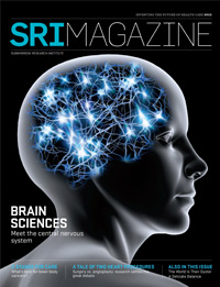
Imaging and the injured brain: detecting the obvious and the insidious

Intracerebral hemorrhage, when a blood vessel inside the brain bursts, either spontaneously or because of trauma, is especially dangerous when it expands further into the brain. Dr. Richard Aviv has discovered the “spot sign,” a marker seen with brain imaging that can predict which hemorrhages are likely to expand.
Photo: Curtis Lantinga
“Stroke is a cataclysmic event,” says Dr. Richard Aviv, a neuroradiologist in the Brain Sciences Research Program at Sunnybrook Research Institute (SRI). The symptoms come on without warning: sudden limb weakness, nausea, slurred speech and loss of vision, among others.
There are several types of stroke, but all disrupt blood flow in the brain and rob its tissues of oxygen, causing neurons to die. Hemorrhagic stroke, or a brain bleed, occurs when a blood vessel suddenly leaks or bursts inside the brain. It is the deadliest type of stroke: about 40% of people who have a hemorrhagic stroke die in hospital, and most survivors are left with disability, which can include paralysis, loss of speech, blindness and dementia.
No effective, targeted therapy to stop the bleeding in a hemorrhagic stroke exists. Hypertension is the most common culprit, and in those cases, blood pressure control can help reduce bleeding risk. With aging, amyloid deposits in the walls of the blood vessels can also give rise to bleeding in the brain. “Alzheimer’s disease, which is associated with amyloid deposits, is becoming a more common cause of cerebral hemorrhage,” says Dr. Sandra Black, director of the Brain Sciences Research Program at SRI. Surgery is only suitable for a few patients.
“There is a huge, unmet need for a safe and effective treatment for brain hemorrhage,” says Dr. David Gladstone, a stroke neurologist in the Brain Sciences Research Program at SRI and director of the Sunnybrook Regional Stroke Prevention Clinic.
The first few hours after an intracerebral hemorrhage occurs are critical. Although some brain bleeds stop quickly and spontaneously, aided by natural pressure within the brain and blood-clotting factors, others continue to bleed, and become fatal or leave a person permanently disabled.
“We see some patients arriving at the emergency room while the brain hemorrhage is still at a small size—they’re awake, talking and moving, and stroke symptoms are relatively mild. However, minutes or hours later, many will be in a coma and in critical condition because of an enlarging hemorrhage,” says Gladstone. “We desperately need a treatment to prevent this catastrophic progression,” he adds.
Until recently, clinicians had no way to predict which hemorrhages would worsen. Then, in 2007, Aviv and colleagues developed the “spot sign,” a noninvasive imaging sign that can identify patients with an intracerebral hemorrhage that will likely grow. It uses an imaging test called a computerized tomography (CT) angiogram to look at blood vessels in the brain. The test relies on a contrast dye to highlight the vessels on the image; the dye will leak out of ruptured vessels and pool nearby, forming a bright white spot in the middle of the hemorrhage on the image.
The spot sign is a mark of active bleeding in the brain. Between 25% and 33% of patients with an intracerebral hemorrhage have one or more spot signs. Because the CT angiogram scan takes only two minutes, the spot sign has enabled clinicians in the emergency department to predict quickly and accurately which patients are likely to get worse.
The more spots there are, the greater the chance the hematoma will expand. “The bottom line is that one is too many,” says Aviv. “There is a lot of optimism amongst neurologists that finding a spot sign may eventually mean a treatment for intracerebral hemorrhage. It’s an orphan disease without a cure, and we hope that we’ve discovered the sign that will allow at least the testing of potential candidates for a cure.”

One such drug has shown promise. Recombinant activated factor VIIa, a blood-clotting drug used in hemophilia, is known to stop bleeding quickly. Gladstone and his colleagues wanted to test it in a subgroup of patients with a spot sign who could benefit most from the drug. Previous studies had not been done with any image-guided techniques to target the therapy to these highest-risk patients.
“We know the spot sign is a marker of bad outcomes, including prolonged hospital stay, death and morbidity,” says Aviv. “Now we want to know: can we intervene when we see this marker to prevent the poor outcome?”
Stopping the leaks
In 2011, Gladstone, Aviv and their colleagues launched SPOTLIGHT, a first-of-its-kind clinical trial of an image-guided emergency treatment protocol for intracerebral hemorrhage. This trial aims to stop brain bleeds from growing bigger by identifying patients with a spot sign and treating them in the emergency room with an injection of factor VIIa. The spot-sign-positive patients—the active bleeders—identified by the imaging scan are thought to be the ones who will respond best. The patients who don’t have a spot sign are unlikely to worsen and therefore are not enrolled.
The study is headquartered at Sunnybrook and involves 12 stroke centres across Canada. They expect it will take about five years. “The hope is that this treatment, if administered quickly enough and to the right patients, might save lives and reduce disability in survivors,” says Gladstone.
Outside the clinic, Aviv is tackling the biology of intracerebral hemorrhage. As is often the case in medicine, researchers and clinicians need to learn more about how a condition, in this case intracerebral stroke, begins and develops. “We don’t know enough about how hematomas grow,” says Aviv. “We wanted to see how hematomas expand, and whether there’s a cascade effect where one vessel ruptures, followed by another, et cetera.”
This is the first preclinical model of the spot sign for acute intracerebral hemorrhage.
Dissatisfied with existing models that aimed to approximate intracerebral hemorrhage, Aviv created a model that more closely mimics acute human hemorrhagic stroke. He worked with Dr. Kullervo Hynynen, director of Physical Sciences at SRI and a pioneer in the development of focused ultrasound, to design a way to rupture the blood vessels in the basal ganglia, a frequent site of hemorrhagic strokes. Using magnetic resonance imaging (MRI) to guide a high-intensity ultrasound wave to the right spot in the brain, they were able to cause a more localized brain bleed than could be done with other models.
This is the first preclinical model of the spot sign for acute intracerebral hemorrhage. Its creation opens up myriad possibilities.
Aviv says they are now able to understand the different phases of hematoma growth, and have a model they can use to study therapies to treat people with an ongoing brain hemorrhage. “We totally think this simulates the human condition,” says Aviv.
Covert attacks
Not all strokes are so evident. Silent strokes lack the obvious symptoms associated with stroke, but still cause damage. They often happen in the brain’s white matter—its inner core. This layer is more vulnerable to disruptions in blood flow because fewer blood vessels traverse it than the cerebral cortex (grey matter). Silent strokes can be hemorrhagic (bleeding) in nature, but they are mainly due to a blockage of the small arterioles that plunge into the white matter.

As a neurologist, Dr. Sandra Black sees first-hand how intricately linked stroke and dementia are—it’s all about the vasculature, she notes.
Photo: Curtis Lantinga
Black, who sees patients with stroke and dementia, says that many patients come to the clinic because they have a memory complaint, or they don’t seem to be functioning as they normally do. Often the family is worried. “The main effect of the small strokes is that they may interfere with the flow of information between the front and the back part of the brain—you get executive dysfunction,” says Black. “With respect to memory, you can encode the information, but you have trouble retrieving it.”
Silent strokes are more common in older people and linked to future problems. A study of 3,660 people with an average age of 75 years revealed that 28% of seniors had silent strokes. Another study that followed-up participants for four years found that those with evidence of past silent strokes were more likely to experience problems with memory and thinking, and develop dementia and stroke.
Black, along with Dr. Brad MacIntosh, a medical biophysicist at SRI, and their colleagues are finding links between silent stroke, white matter disease and dementia. (See Infrastructure Issues.) They’ve used a functional MRI technique to uncover evidence of silent strokes and white matter disease, and understand their origins and impact on the brain and its function. The technique shows bright white spots where silent strokes have occurred and diffuse white areas, called white matter hyperintensities.
The common factor between the three—silent stroke, white matter disease and dementia—is the vasculature that feeds the brain and removes its waste. The white spots and larger areas are signs of reduced blood flow, which can have knock-on effects. For example, if pathways along the vasculature can’t clear out dangerous proteins like beta-amyloid, which damages cells, then amyloid deposits may form within the brain, and the person may develop dementia. “The vasculature is intimately related to the development of Alzheimer’s disease. But it also works in the other direction: Amyloid deposits can cause blockages and hemorrhagic stroke,” says Black.
Black and MacIntosh expect that by understanding the early changes to the brain’s blood vessels, intervention before people succumb to stroke, cognitive decline or dementia might be possible. People at risk can try to reduce their vascular risk factors by keeping hypertension under control with diet, exercise and medications prescribed by their physicians. “You need to do activities that increase brain blood flow and make it healthier,” says Black.
“Some people say that having silent strokes is a part of getting old, and that we should just move on,” says MacIntosh. “I think that’s a pessimistic point of view. We should be chipping away at this attitude, so that we can age as best as we can by preserving our brain’s health.”
Research Funding and Other Information
Aviv (excluding SPOTLIGHT trial): Canadian Stroke Consortium, Heart and Stroke Foundation of Ontario and NovoNordisk Canada. Black: Alzheimer Society of Canada, Canada Foundation for Innovation (CFI), Canadian Institutes of Health Research (CIHR), Heart and Stroke Foundation Canadian Partnership for Stroke Recovery (CPSR), LC Campbell Foundation, and the Ontario Ministry of Research and Innovation (MRI). MacIntosh: CIHR, CPSR, and Natural Sciences and Engineering Research Council. SPOTLIGHT trial (Aviv and Gladstone): CIHR, MRI and Ontario Stroke Network. Aviv is a faculty member within the department of medical imaging at the University of Toronto; Black and Gladstone, medicine (neurology); MacIntosh, medical biophysics.


