Cover Slip

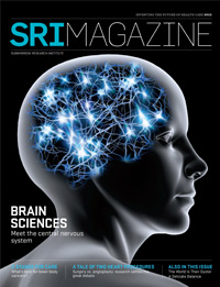
Image: taken with a Zeiss LSM510 confocal microscope. Labelling of the brain sections was done by Lillian Weng; imaging by Dr. Emmanuel Thévenot; selection, Jonathan Oore. Dr. Isabelle Aubert did the post-processing.
In the brain, amyloid plaques are a hallmark of Alzheimer’s disease. When focused ultrasound is delivered through the skull, the activation of astrocytes (star-shaped red cells) and microglia (blue) could contribute to reducing the number and size of amyloid plaques (green). Biologists and physicists at Sunnybrook Research Institute have joined forces to investigate these processes.

