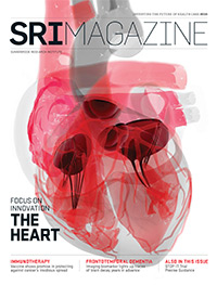
A Scar Runs Through It
June 15, 2015

Dr. Graham Wright and colleagues at Sunnybrook Research Institute have optimized magnetic resonance imaging techniques to identify different types of scar tissue in the heart.
Photo: Doug Nicholson
Scars tell stories of battles fought and won. For many people, they are a source of pride, but when it comes to the heart, these scars are often a harbinger of troubles to come.
During a heart attack, blood flow to the heart is restricted, leading to a shortage of oxygen to the muscle. Without oxygen, cells in the myocardium, the muscular tissue of the heart, die. The heart tries to repair itself by replacing the dead tissue with scar tissue, which is often hard and fibrous. Although scar tissue helps to maintain the structural integrity of the heart muscle, the cardiac fibroblast cells comprising the scar cannot beat like healthy heart muscle cells. The combination of increased hardening of the heart muscle and reduced numbers of beating cardiac muscle cells diminishes the blood-pumping ability of the heart. Over time, this scar tissue can disrupt the normal electrical signals in the heart, leading to abnormal heart rhythms and, eventually, heart failure.
Dr. Graham Wright, director of the Schulich Heart Research Program at Sunnybrook Research Institute, has pioneered the use of magnetic resonance imaging (MRI) to detect and characterize scar tissue in the heart. Using preclinical models, he and his team have developed and optimized MRI methods to identify healthy tissue, scar tissue and the border zone in-between, which contains a mixed population of healthy cardiac muscle cells and scar fibroblast cells. The heterogeneous composition of cells in the border zone imparts unstable electrical properties to the area, making it a fertile breeding ground for arrhythmias, or abnormal heart rhythms.
In two studies published in 2013, Wright along with colleagues Drs. Eugene Crystal, Nilesh Ghugre and Greg Stanisz showed that MRI can determine the extent of the border zone and scar. Moreover, they showed that MRI can complement electrophysiology tests in accurately pinpointing arrhythmia-prone areas. Knowing the location and size of scar tissue helps doctors predict which patients will develop potentially fatal arrhythmias and administer more targeted therapies.
Recent efforts to mend the broken heart have focused on stem cells, which have the potential to become any cell type. The goal of stem cell therapy is to regenerate healthy cardiac muscle cells after a heart attack to replace the dead tissue. Preliminary studies in patients and preclinical models have demonstrated great promise—stem cell transplantations following a heart attack reduced scar tissue formation, increased heart muscle regeneration and improved cardiac function.

