Projects
The projects in the Breast Cancer Research Centre (BCRC) require multidisciplinary collaboration among scientists, their lab groups and clinical professionals. These projects position basic scientists and clinical trials experts in close proximity to patients, as researchers work on the development and implementation of sophisticated imaging techniques and genetic tests to help make surgeries and prognoses more accurate. Ultimately, research in the centre aims to make the process of care more seamless for breast cancer patients by merging basic science with clinical application to develop tailored treatment options.
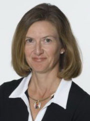 |
Claire Holloway Dr. Holloway's work with Dr. Donald Plewes and Dr. Martin Yaffe is enabling more accurate breast imaging before and during surgery (via magnetic resonance imaging [MRI] and ultrasound, respectively), and providing more detailed knowledge after surgery on whether all cancerous tissue has been removed. Tumours extracted during surgery lose their form and outline; Holloway is developing a better method to determine if all cancerous tissue has been removed by maintaining a specimen's shape, testing its edges and combining special imaging techniques, such as MRI, with new specimen-processing methods. The centre's high-tech experimental operating room is essential to the work of Holloway, an associate scientist at SRI, and has allowed her to perform more than 150 surgical procedures. This research will make breast cancer surgery more precise. |
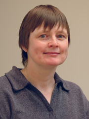 |
Anne Martel Contrast-enhanced breast imaging techniques including MRI, computed tomography and ultrasound are becoming more complex, making interpretation of the increasing amount of information they provide more difficult. Dr. Martel, a scientist at SRI, and her lab are developing new computational techniques to identify the clinically relevant information delivered by these new imaging tools. The resulting parametric maps and classification images enable clinicians to make clearer diagnoses of malignant and benign breast lesions. Dr. Martel is also working to improve the quality of breast image data by correcting for patient motion algorithmically and reducing the effects of artifacts and noise. |
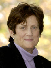 |
Kathleen Pritchard Dr. Pritchard's goal is to see all women with breast cancer treated with drugs highly specific to their cancer, based on their genetic and hormonal makeup, instead of with chemotherapy, which often has debilitating side effects. She has led and contributed to many practice-changing trials. For example, she co-led a trial of letrozole, a drug with fewer side effects, to help stop the cancer from returning in postmenopausal women who had already taken tamoxifen for five years. The drug's effects were dramatic, reducing the risk of recurrence more than 40%. This led to a Health Canada approval for the therapy in 2005. With senior scientist Dr. Arun Seth and SRI associate scientist Dr. Wedad Hanna, Pritchard is also working to expand the Henrietta Banting tumour/tissue databank, a warehouse of information on a retrospective cohort of 1800 women treated surgically for breast cancer first between 1977 and 1987, and then between 1987 and 1997. Pritchard and her colleagues have compiled information on recurrence and outcomes, and have prepared tissue microarrays for more than 5800 of the women. The aim of this project is to develop molecular methods, via gene profiling, to predict disease outcome and tailor individualized treatments. The database is an integrated resource for use across Canada by researchers who seek to validate potentially useful new molecular markers. |
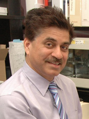 |
Arun Seth The primary aim of Dr. Seth's BCRC research is to understand the molecular mechanisms of breast cancer progression and metastasis. His lab uses novel mouse models and molecular approaches to develop therapeutic strategies that will interfere with the function of key proteins that affect breast cancer. Seth and his lab have isolated a number of genes associated with the ubiquitin-mediated protein degradation pathway. This pathway plays a major role in the degradation of many proteins involved in the cell cycle, proliferation, apoptosis and progression and metastasis of breast cancer. Seth is also the director of SRI's BCRC-linked genomics facility, which allows for fingerprinting of the genes associated with individual tumours. Pure cell populations are isolated from other tissue, enabling study of the key molecular events in the progression of breast cancer through comparison of gene expression in normal tissue, noninvasive and invasive cancers, and cancers that have spread to other locations in the body. |
 |
Martin Yaffe Dr. Yaffe, a senior scientist at SRI, and his basic physics lab develop technology to improve breast cancer detection, diagnosis and treatment. Yaffe's work ranges from design, fabrication and evaluation of high-resolution, solid-state imaging sensors for digital mammography, to development of image-processing strategies for improved diagnostic image quality, to 3-D imaging (tomosynthesis), contrast-enhanced imaging methods and, most recently, development of a system for 3-D imaging of breast histology to help improve the accuracy of breast surgery. Yaffe, in collaboration with SRI associate scientist Dr. Roberta Jong, played a major role in DMIST, an international trial that compared the effectiveness of standard mammography to digital mammography in close to 50,000 women, and for which Sunnybrook was the sole Canadian site, enrolling 2500 women. The results showed that for most women the two kinds of mammography work equally well. For certain women, however—those who are premenopausal, younger than 50 or who have dense breasts—digital mammography is more sensitive, and thus detects tumours more accurately. These results had practice-changing implications. |


