Enmeshed in research
By Eleni Kanavas
Amani Ibrahim has always been interested in the medical field. In high school, she thought she would go to university, study life sciences and become a doctor. Instead, she found herself gravitating toward another academic path in computer sciences and technology.
In May, Ibrahim completed her four-year undergraduate degree with honours in computing and mathematics from Queen’s University in Kingston. While working in the “perk lab,” a facility for percutaneous surgery at Queen’s, she learned about a summer research opportunity in the orthopaedic biomechanics laboratory (OBL) at Sunnybrook Research Institute (SRI) led by Dr. Cari Whyne, director of the Holland Musculoskeletal Research Program.
“My professor said there was an opportunity [at SRI] with Dr. Whyne and that they needed a student who was familiar with 3-D Slicer, a free, open-source software platform for the analysis and visualization of medical images, and for research in image-guided therapy,” she says. It was the same program that she uses in her lab in Kingston, so she decided to apply. “I got in contact with Dr. Michael Hardisty, who works under Dr. Whyne, and it was a good match.”
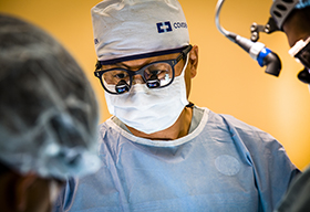
Ibrahim is working on a project that is part of the OBL’s collaboration with Calavera Surgical Design Inc., a startup company out of SRI that produces custom craniofacial implants for patients with facial trauma. Calavera has developed a low-cost surgical process that uses patient-specific molds generated from computed tomography (CT) data to bend mesh for surgical implants. The implants are used to restore normal anatomy in the skull and facial bones.
Her project focuses on developing software to improve the image-processing technology used by Calavera in their design of custom implants for thin bone structures. Computed tomography imaging for fine boney structures such as those around the eye sockets have been a particularly difficult and time-intensive design challenge because the resolution of the CT scans is low.
“I’m working in collaboration with Calavera, so I am writing code for Dr. Hardisty. I am also learning about and utilizing existing image-processing algorithms, which are of course mathematically based,” she says. “The main image-processing algorithms I use are in the form of filters for sharpening and deblurring images, and creating smooth continuous surfaces from point data.”
She notes the main issue with CT scans is that they do not register areas of fine bone very well. This means they appear to be blurry in imaging programs, as if there are holes in the 3-D model.
“My job is to basically fill in those holes or use imaging tools to make those holes nonexistent in the first place. It’s been interesting to find a way to do that,” she says. “I’ve just been reading about and testing image-processing techniques that could be useful for image enhancement and model hole-filling.”
Ibrahim is also designing a streamlined workflow of the software plans, so that they are user-friendlier. Her day-to-day schedule fluctuates between working on the modules of the “plan creation” workflow. This involves creating the different steps and modules to make sure they work independently and flow well together, as well as ensuring the workflow as a whole creates a good user experience, she explains.
The steps include loading CT data into the program and improving the quality of the CT scans by deblurring or using other image-processing techniques. This is followed by converting the scans from CT volumes to 3-D models, filling in any remaining holes in the model, and registering and clipping the model. Finally, the scan is exported into a new 3-D model design.
“When this is done successfully, it creates a much sharper image where the fine bone structures no longer look like gaps,” she says.
Immersed in a group of researchers from clinical, engineering and medical physics backgrounds, she says she enjoys working in such a collaborative and supportive environment that includes an interesting mix of research and software development.
When asked what has been the most rewarding part of working in the Whyne lab, Ibrahim says, “the freedom to be able to produce the best work I can.” Next month she will be starting a master’s degree in biomedical computing with a focus on image processing at Queen’s University.
“As a programmer, the software I am currently working on has been one of the largest projects I have built so far. It has really helped reinforce the idea of writing efficient modular and reliable code, and keeping communication lines open with my amazing team,” she says. “I think [that] overall, it has taught me how to be a better researcher and learner, both [of which] fundamental for succeeding in my master's.”
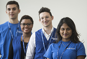
Amani Ibrahim is part of the D+H SRI Summer Student Research Program.


