Canada Research Chair awarded to leading heart scientist
By Jim Oldfield
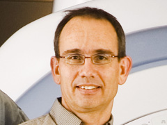
Dr. Graham Wright has been awarded the Tier 1 Canada Research Chair (CRC) in Imaging for Cardiovascular Therapeutics.
"It's a recognition of our research in imaging for cardiovascular therapeutics, and of the people doing that work," Wright says. "We have a lab of 20 to 30 people working as a team with a lot of ideas, and it's very gratifying that the scientific community thinks it's worthwhile."
Wright is the director of and senior scientist in the Schulich Heart Research Program at Sunnybrook Research Institute (SRI), and a professor of medical biophysics at the University of Toronto. He is also a scientist in SRI’s Centre for Research in Image-Guided Therapeutics.
The award guarantees $1.4 million in salary over seven years. The validation that comes with the award—proposals are peer-reviewed by Canadian and international researchers after nominations by each chair-holder's home institution, in this case SRI, and subsequently U of T—should provide an important boost, Wright says, to the collaboration and institutional support that is crucial to his translational research.
Team boost
"Imaging for cardiovascular therapeutics is not research where I can work alone, focusing only on theory. It's a big venture, and it needs a multidisciplinary team pulling together in the same direction with substantial resources," says Wright. "Such a diverse group needs validation that what they're doing makes sense, because it's a long road to bring this work to fruition." The award provides the stability to take on such long-range projects.
Early in his career, Wright studied a functional magnetic resonance imaging (MRI) issue—oxygen levels in blood—and he pioneered the first method for noninvasive, in vivo measurement of blood oxygen. Clinicians are testing this technique in the diagnosis and assessment of heart conditions.
Better diagnosis and treatment
A logical evolution of this work is linking the powers of diagnostic imaging more closely with therapy. His research program has evolved toward imaging physiology precisely and rapidly within a procedure suite, in hopes of making more accurate treatment decisions and directly guiding cardiac interventions.
In particular, Wright and his team will study quantitative measurement of myocardial tissue properties with MRI, develop new image analysis and visualization tools, integrate catheter-based sensing for local measurement during MRI-guided interventions, and demonstrate the efficacy of new imaging tools and therapies in preclinical models and patients with occlusive vascular disease and other heart conditions.
The need for improved and more cost-effective ways to diagnose and manage cardiac conditions is urgent. Cardiovascular disease is the leading cause of death in North America; in Canada, it accounted for more than 69,000 deaths in 2006. Owing to advances in detection and treatment, however, death rates from heart disease and stroke dropped about 30% in Canada between 1994 and 2004. This decline has created further need for better chronic disease management.
Collaboration essential
In addressing that issue, Wright's research is necessarily cooperative—he works with engineers, image analysts, computer programmers, physiologists and clinicians, as well as researchers investigating complementary modalities like X-ray and ultrasound.
He says he is committed to fostering and building on those collaborations: "Close ties with the clinical program make the work more rewarding, because we're striving toward solutions that are going to make a difference to people. It's critical to work with clinicians and their patients so we can make sure we're answering the right questions."


