The impact of metastatic disease on vertebral bone quality
Bone quality is important from mechanics and material perspectives when considering vertebral structural integrity. Quality is dependent on mechanical (material and structural) properties, including mineralization, composition, remodelling, connectivity and architecture. To understand how metastatic disease (osteolytic and osteoblastic) affects bone quality we developed preclinical nude rat models of Osteolytic (HeLa), Mixed (ACE-1) and Osteoblastic (ZR-75) metastases. These models highlighted changes in the properties of both the mineral (hydroxyapatite) and organic (collagen) phases of the metastatic bone tissue, which correlated to tissue level material behaviour and whole vertebral mechanical properties. Computationally we have developed algorithms to directly measure deformation and strain via registration of loaded and unloaded µCT scans.
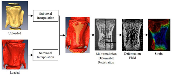 Multiresolution Deformable Image Registration Pipeline
Click to view plain text of infographic
- Unloaded, Subvoxel Interpolation
- Loaded, Subvoxel Interpolation
- Multiresolution Deformable Registration
- Deformation field
- Strain
Load and boundary conditions from this experimental imaging data have been applied to continuum and micro finite element (µFE) models of healthy and metastatically involved vertebrae. Microdamage present in healthy and metastatically involved vertebrae can be identified histologically through fuchsin staining, chelating agents and barium sulphate staining combined with µCT imaging. For higher resolution information, back scatter electron microscopy can be performed on BaSO4 stained sections. µFE models of these vertebrae were able to represent the load induced microdamage seen in these images. Microdamage was found to be spatially correlated with regions of high stress and strain in our µFE models with clear microdamage thresholds identified. Yet mechanically induced microdamage thresholds were different in healthy vs metastatic bone suggesting tissue level changes in bone quality.
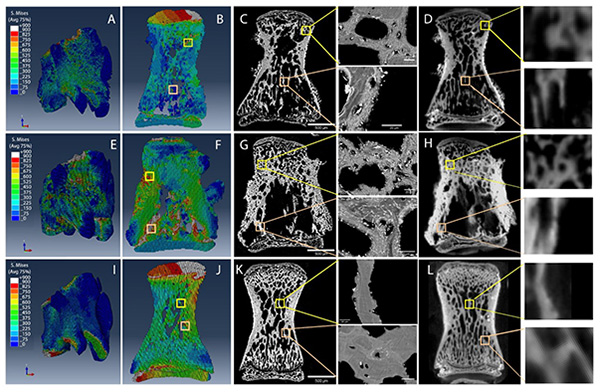 Micro Finite Element Modelling, Backscatter Electron Imaging and µCT imaging of BaSO4 stained Osteolytic (top), Mixed (middle) and Control (bottom) vertebrae.
Recent work has gone beyond modelling of stress and strain, demonstrating the ability of non-linear FE models incorporating cohesive elements to represent post-yield modeling of crack initiation, propagation and failure. The impact of multimodal treatments on bone quality in the metastatic skeleton is an ongoing area of investigation.
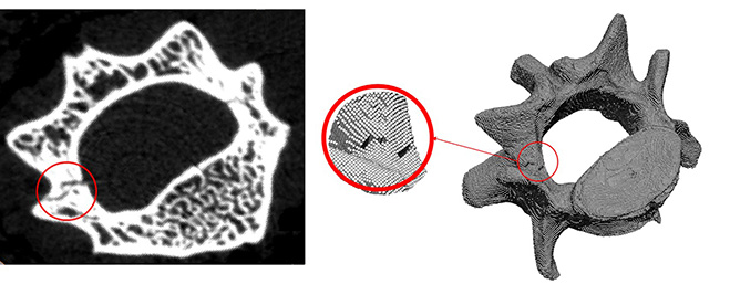 Micro FE vertebral model generated from microCT data incorporating cohesive elements to enable the modelling of post yield behaviour/fracture.

