About the biomarker imaging research laboratory
The biomarker imaging research laboratory is a cutting-edge research facility that develops new histopathology techniques and image-processing tools to enable imaging pathology correlation analyses in cancer and other diseases. It is a fundamental resource within the Centre for Research in Image-Guided Therapeutics.
One main focus of the lab is biomarker panel development and validation using multiplexing technology, molecular profiling and multimodality image-processing algorithms for digital pathology and in vivo medical imaging.
The lab uses an immuno-fluorescence multiplexing digital imaging system developed by the General Electric Global Research Center for validating reliable quantitative prognostic and predictive biomarkers. The system is being used to characterize cancers and their environment to provide more precision therapies.
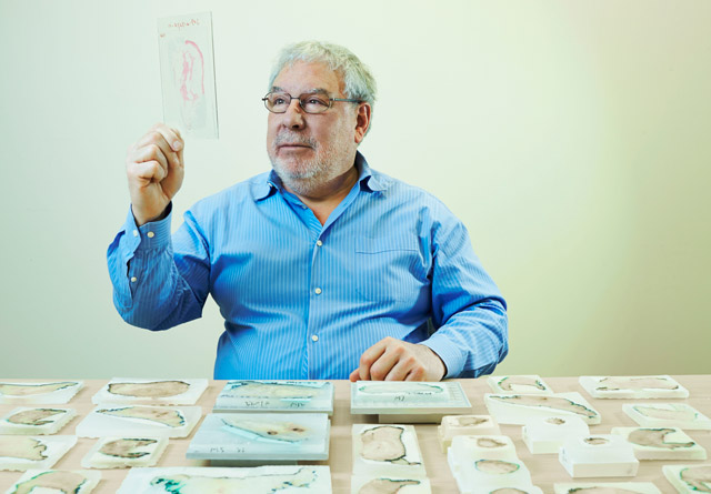
The lab operates under the licence and supervision of the department of anatomic pathology at Sunnybrook to produce large, whole organ diagnostic slides through an alternative tissue-processing method called whole mount. It has developed a full range of digital whole-mount histopathology techniques that produce large (up to 5” x 7”) tissue slides from breast, prostate, tongue, colorectal and brain cancer tissue samples.
It has implemented clinical standards for quality control and process monitoring, including clinically compatible tissue tracking and sample storage systems. Several state-of-the-art digitizers are available to accommodate various sample sizes, and to perform bright field and fluorescence scanning of regular and large whole-mount slides.
In collaboration with other labs, our work is focused on quantitative 3-D histopathology, including automated image processing and computer detection of structures.
Principal investigator
Dr. Martin Yaffe
View profile
Contact
For service and quotations please contact:
Yulia Yerofeyeva
Program manager, Ontario Institute of Cancer Research/BIRL
2075 Bayview Ave.
Room C7 50a
Toronto, ON M4N 3M5
416-480-4265
yulia.yerofeyeva@
sri.utoronto.ca

