Whole-mount histopathology
The biomarker imaging research lab specializes in whole-mount histopathology, which captures an entire cross-section of a surgical tissue specimen. These tissue sections are up to 120mm x 170mm in size and, once stained and scanned, can be used in imaging-pathology correlative studies.
Whole-mount histopathology provides improved sampling through preserving spatial arrangement of the tissue, which is key for studying disease characterization and progression such as spatial heterogeneity and progression of cancer. Other advantages of this technique are:
- more accurate surgical margin assessment;
- volumetric disease burden; and
- secondary disease foci identification
Over the years the lab has processed a large variety of whole mount tissue samples: breast cancer (lumpectomies and mastectomies), whole mount prostate cancer, oral (tongue) cancer, colon cancer, and lately, pancreas, brain and various arteries.
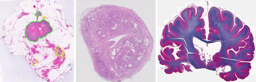 Left: breast cancer lumpectomy with multi-focal cancer, middle: prostate cancer, right: brain whole-specimen section for neurological research.
These large and regular tissue slides can be digitized in our lab using large field-of-view microscopes and tissue scanners such as Tissuescope LE. Tissuescope can produce high-resolution (25X and 50X) digital images of the slides, which can be converted into 2D and 3D datasets.
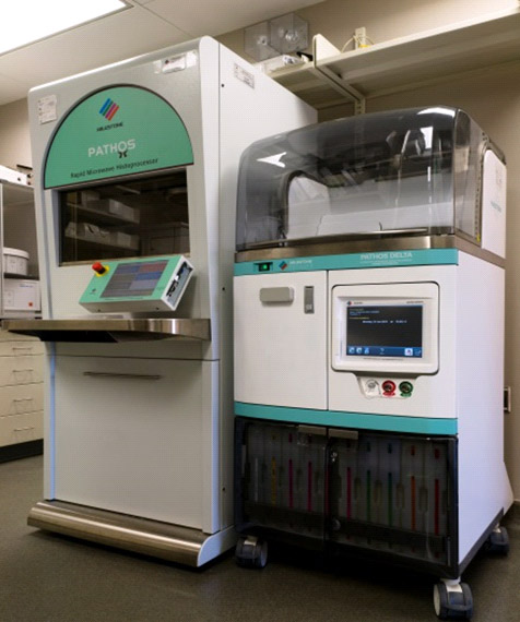
We are continuing our work on the improvement of the whole-mount process by optimizing laboratory procedures, using advanced equipment and applying automation to the workflow.
We have developed a prototype automated stainer for whole-mount slides to reduce further the cost and time associated with the method, and validated it on several clinical whole-mount hematoxylin and eosin slides.
Examples of automation include prototype automated stainer, automated microwave processing methods that significantly reduces overall processing time.
A Tissue slicer was developed and patented for improved sampling of large tissues and has been used routinely to render accurate slicing of the specimens to produce serial sections at equal intervals.
To accurately correlate pathology and in vivo imaging, we have developed an alternative gel formulation that allows rapid embedding and accurate slicing and sampling of the tissue. providing a multitude of opportunities for radiomic studies.
 Tissue processing equipment (click to enlarge)
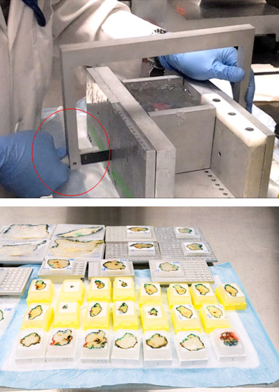 Tissue slicer (click to enlarge)
Tissue processing equipment
Tissue slicer

