Image processing and Finite Element modelling of the CMFS
Deblurring clinical CT scans to restore thin bone structures
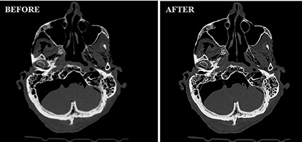
In clinical CT imaging, there are severe errors in geometry and material properties in thin bone structures, which are in large part due to blurring. As such we aimed to restore sub-mm bone geometry and intensity information in clinical CT images by post-reconstruction image processing. Knowing what the non-blurred profile of cortical bone should look like, we can estimate it based on the observed profile in low-resolution CT images to restore the 3D anatomy of the craniomaxillofacial skeleton (CMFS).
Finite Element modelling of the CMFS
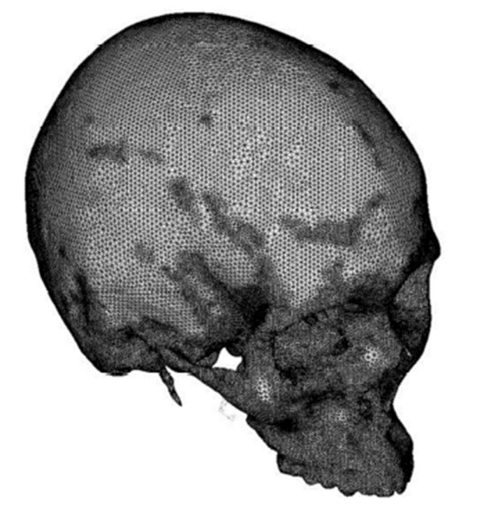 Computed tomography (CT) 3D geometrical reconstruction of the skull.
We created a new pipeline to generate accurate Finite Element (FE) models of the CMFS with heterogenous application of bone modulus values based on deblurred CT image intensity. Due to the very thin nature of the bone, this used nodal based assignments and algorithms to deal with partial volume effects at surfaces.
Mechanical testing is performed on specimens instrumented with strain gages to validate our modelling. Under more physiologic loading conditions, these models are able to show important pathways of load transmission occurring through the complex areas of the zygoma and orbital rim.
Understanding the sensitivity to load and boundary conditions, we are employing new methods to better represent muscle loading. MR-based diffusion tensor imaging (DTI), in combination with anatomical data, can elucidate the fibres and fibre bundles associated with the muscles of mastication. We are concurrently developing models focused on representing loading post-fracture reconstruction towards optimizing hardware requirements for stable fixation.
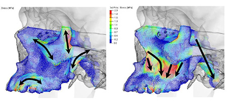 Load transmission through the zygoma and orbital rim.
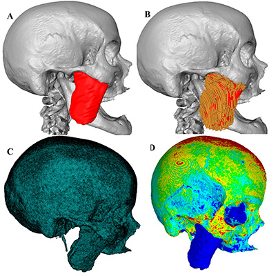 A) Masseter and skull volumes obtained from MRI and CT, B) Masseter fiber architecture obtained from DTI within the masseter volume, C) Mesh generation of the skull and masseter, D) Heterogeneous distribution of bone elastic modulus obtained using CT-based bone densities.
