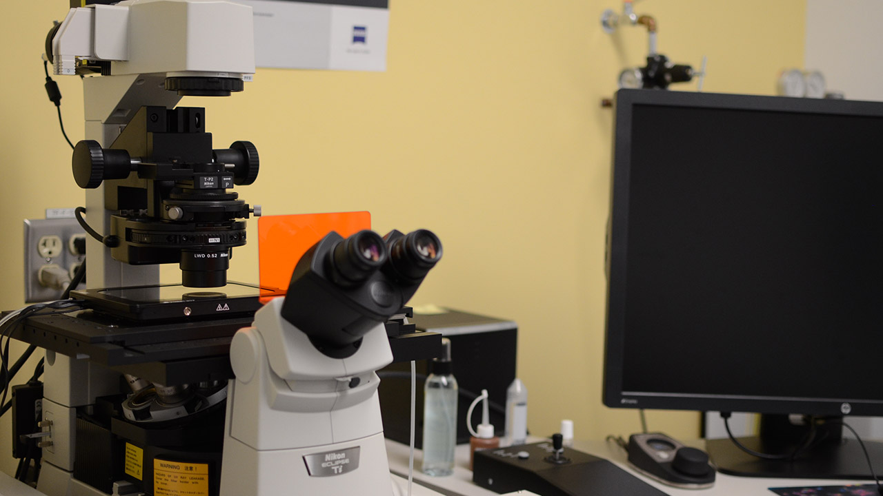
Tool kit: Nikon A1 laser scanning confocal microscope
Sunnybrook Research Institute (SRI) has upgraded its microscopy facilities with the addition of the Nikon A1 laser scanning confocal microscope. “The main advantage is that you can get really great resolution,” says Dr. Geneve Awong, manager of the Centre for Flow Cytometry and Microscopy where the new microscope is housed.
The Nikon A1 is equipped with new scanner and image correction technologies that enable users to acquire high-resolution images of up to 4096 x 4096 pixels. It is also capable of high-speed acquisition at 10 frames per second for 512 x 512 pixel images, which allows researchers to observe dynamic processes in real time. Other unique features of the microscope include a 32-channel spectral detection to identify fluorophores with overlapping emission spectra; a high-content analysis program for high-throughput imaging; and a perfect focus image stabilization system. “The perfect focus system allows you to autocorrect and keep that nice focus through the whole well and in all wells during imaging,” says Awong.
Dr. Isabelle Aubert, a senior scientist in the Hurvitz Brain Sciences Research Program, is one of the main users of the new microscope. “The quality of the image that you can get for fluorescence microscopy is significantly improved from a spinning disk,” says Danielle Weber-Adrian, a PhD student in the Aubert lab. Weber-Adrian is studying noninvasive gene delivery to the brain using focused ultrasound (FUS). Following FUS treatment, she uses the Nikon A1 to image thick sections of the brain and look for fluorescent signals from her therapeutic. “I want to know how much got into the brain and where did it go,” she says. The powerful new microscope enables her to determine not just where in the brain her therapeutic is, but specifically in what cell types and what areas within a cell. Prior to SRI obtaining the Nikon A1, Weber-Adrian had to travel downtown to use a laser scanning confocal microscope. “Now I don’t have to take the shuttle downtown [with my samples],” she says. “This is a huge improvement.”
Other SRI researchers who will use the laser scanning confocal microscope include Drs. Kullervo Hynynen, Marc Jeschke and Burton Yang. Dr. Rob Screaton, a senior scientist in Biological Sciences, will also be using it to conduct imaging screens to identify new genes involved in pancreatic beta cell and mitochondrial biology.
The Nikon A1 laser scanning confocal microscope is worth $630,000 and was purchased with funding from the Canada Foundation for Innovation.