A sound pairing
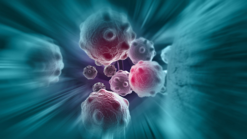
“No man is an island,” declared the British poet John Donne, speaking to the interdependence of people. Cancer cells behave similarly in that they rely on neighbouring blood vessels, immune cells and other healthy tissue to survive. This supportive network, referred to as the tumour microenvironment, is vital to the growth and spread of cancer cells. For instance, blood vessels deliver oxygen and nutrients, while wound-healing cells called fibroblasts provide structural support. A sticky network of molecules, known as the matrix, supports all the cells and binds them together.
Monitoring a tumour’s immediate surroundings can provide important clues about the status of a cancer in response to treatment, says Dr. Stuart Foster, an imaging physicist at Sunnybrook Research Institute (SRI). “In response to adequate blood supply and the right molecular signals there’s a lot of opportunity for the tumour cells to respond by multiplying and dividing and growing, as tumours do. On the other hand, if you have an effective therapy, you could slow down the transformation of the microenvironment, restricting the tumour’s ability to develop and spread.”
Foster and colleagues have found that a combination of two techniques, contrast ultrasound and photoacoustic imaging, offers a window into a tumour’s microenvironment noninvasively. Contrast ultrasound involves injecting gas-filled particles into the circulation, which report signals back to the ultrasound scanner to show how blood is flowing through the tiny blood vessels in tissue. Photoacoustic imaging uses short laser pulses to excite ultrasonic signals detectable by ultrasound. By choosing very specific wavelengths it is possible to translate this information into maps of tissue oxygenation.
In a paper published in Cancer Research in August 2016, the team demonstrated for the first time that combining the techniques yielded detailed pictures of blood flow and oxygenation in tumours in preclinical models of colon, prostate and breast cancer. These images depicted the structure of blood vessels feeding the tumour, how well they worked and whether they were leaky. The researchers validated their findings by analyzing tumour samples microscopically.
Tumours that don’t get enough oxygen are said to be hypoxic, and they represent an aggressive type of cancer. Unlike normal cells, which die under extreme oxygen deprivation, cancer cells harbour genetic mutations that enable them to adapt to and thrive in this harsh environment. Moreover, low oxygen levels in tumours render radiation therapy and chemotherapy less effective. Thus, a tumour’s oxygen level could serve as a biomarker to help doctors determine a patient’s prognosis, says Foster, adding, “It would be very useful to be able to monitor that situation in a dynamic way with a simple imaging technology like ultrasound.”
Doubly good
In another study published in the same issue of Cancer Research, Dr. Bob Kerbel, a cancer biologist at SRI, used the imaging techniques developed in Foster’s lab to study the effects of pairing two drugs in preclinical models of breast cancer. The first drug, Avastin, is an antiangiogenic drug; it works by cutting off a tumour’s blood supply; however, it has shown limited longer-term effectiveness because tumours develop resistance to it. The second, CRLX101, is an experimental drug that packages a chemotherapy called camptothecin in a nanoparticle. The latter suppresses a protein called hypoxia-inducible factor 1 (HIF1), which activates genes that help cancer cells adapt to low oxygen levels.
“What Bob is looking at is how to make [Avastin] more effective,” says Foster. “So he combined it with this new drug and asked ‘What happens to the microcirculation [circulation within tumours] when I apply these two drugs together?’ You give the antiangiogenic drug, [and] it shuts down the microcirculation in the tumour, which we can see quite clearly using contrast ultrasound. When you give the second drug, it actually helps circumvent some of the resistance mechanisms that occur when patients take [Avastin] alone.”
The researchers found CRLX101 suppressed the HIF1 protein and concluded that pairing it with Avastin might be a promising approach. The researchers found the drug combination shrank tumours, reduced the cancer’s spread and extended survival in mice. Interestingly, CRLX101 improved blood circulation within the tumour, which, although it feeds the tumour, is also desirable given that it gets more of the drug into the tumour.
“It’s one of the great conundrums of molecular therapeutics of cancer,” says Foster, speaking to the problem of insufficient blood flow in tumours. “People don’t really understand what the best balance is. In this study, which is done in mice, admittedly, the combination of the two [drugs] worked far better than either separately. We could establish that that new drug definitely increased the flow of blood into the tumour, and improved the outcome for the mouse.”
Foster and Kerbel plan to do further studies using ultrasound to study how cancer therapies that exploit the immune system to fight the disease affect the microcirculation of tumours. Foster says he is pleased to be a part of this interdisciplinary research: “It’s an example of a good combination of physics, imaging and biology that helps cancer researchers find answers to interesting questions they’re asking.”