Doors open to new biomedical imaging research suite
By Eleni Kanavas
Sunnybrook Research Institute (SRI) officially opened its biomedical imaging research suite on November 10, 2011. The event was part of SRI's second annual research day for the Centre for Research in Image-Guided Therapeutics (CeRIGT).
"Today we celebrate another milestone in CeRIGT's development," said Dr. Michael Julius, vice-president of research at Sunnybrook Health Sciences Centre and SRI. "The biomedical imaging research suite is unique in its breadth of equipment and multidisciplinary focus, and its integration of preclinical and clinical modalities."
The biomedical imaging research suite is a core facility of CeRIGT in which teams of scientists and clinician-scientists are conducting studies using computed tomography (CT), magnetic resonance imaging (MRI) and focused ultrasound. Their aim is to develop and optimize noninvasive imaging methods for brain, cardiac, cancer and musculoskeletal applications.
"With this facility, we now have the infrastructure to do MR-guided and CT-guided interventions, either clinically or to test new therapeutics," said Dr. Kullervo Hynynen, director of physical sciences at SRI and project lead of CeRIGT.
Hynynen also highlighted the importance of partnerships; SRI's industry partners include Bruker, GE Healthcare, InSightec, Philips and Toshiba, among others.
As part of the celebration, guests enjoyed a cake reception and a tour of the facility.
Cutting-edge equipment
Caron Murray, a senior MRI research technologist at SRI, showed guests the Toshiba Aquilion ONE. The $2.9-million system is a dynamic-volume, whole-body CT scanner with a 320-row detector that can image up to 16 centimeters of anatomy–covering a heart or head–in a single scan. With its high-resolution 3-D imaging, researchers can see how blood flow is moving around an organ, thus leading to better diagnoses in CT angiography. Dr. Graham Wright, director of the Schulich Heart Research Program at SRI, and Dr. Alan Moody, a clinician-scientist and radiologist-in-chief at Sunnybrook, will use the system for diagnostic and therapeutic cardiac studies. Hynynen will use it to plan and monitor focused ultrasound therapies for neurological diseases.
Translating preclinical research into clinical trials
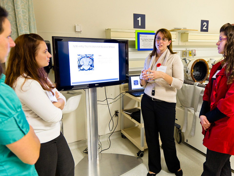
Dr. Alison Burgess, a research associate in Hynynen's focused ultrasound lab, described how the InSightec ExAblate 4000 Brain System could be used in the future to treat patients with stroke.
"Our preclinical work is ongoing as we investigate the optimal HIFU [high-intensity focused ultrasound] treatment of ischemic stroke," she said. "Using our system, we have demonstrated for the first time that it is possible to break up a clot in the brain using HIFU as a standalone method, [but] we have more experiments to perform before being ready for a clinical trial."
Researchers have used the system for preclinical research studies on noninvasive, transcranial MRI-guided focused ultrasound therapy, including blood-brain barrier disruption, said Dr. Yuexi Huang, a senior research physicist in Hynynen's lab. The system was recently approved for clinical trials to treat patients with brain tumours and tremors. "Hopefully, we will treat the first patient very soon," said Huang. Clinician-scientists Drs. Todd Mainprize and Michael Schwartz will lead the clinical trials.
Innovative MRI-guided systems
Dr. Greg Stanisz, a senior scientist in physical sciences, described how the facility will be used across research programs at SRI. "We have five MRI machines dedicated to research that allow us to image everything from cells, to large preclinical models and humans in a variety of different applications ranging from cancer, to cardiovascular to musculoskeletal," he said.
Stanisz is using the Bruker 7-Tesla (7T) MRI system to develop preclinical imaging techniques. This $3.2-million system offers unprecedented sensitivity and throughput-it can image preclinical models at a higher resolution than can any clinical scanner.
Dozens of SRI researchers are already using the 7T system. Experiments include evaluating changes in the vascular and cellular organization of tumours undergoing anticancer therapies; imaging for viral drug delivery in spinal cord injuries; developing nanostructures as contrast agents; and doing behavourial studies in new models of stroke.
Other devices in the biomedical imaging research suite are a GE 1.5T Signa MRI system; a GE 3T long-bore, whole-body, research MRI system; and a C13 dynamic nuclear polarization polarizer for MR metabolic imaging. Sunnybrook Research Institute and The University of Western Ontario are the only institutions in Canada that have a hyperpolarizer system.
The $160-million CeRIGT was established through the Canada Foundation for Innovation's Research Hospital Fund in 2008. The whole of CeRIGT's completion is scheduled for April 2012.
To read more about SRI's second annual CeRIGT Research Day, read this story.


