Research Services
We offer a wide range of immunohistochemistry services for clinical and preclinical research projects and clinical trials. These services are available to internal and external investigators, clinicians, and industry collaborators and investigators. We are committed to exploring new ideas and technologies and welcome new collaborations.

View a plain-text version of infographic
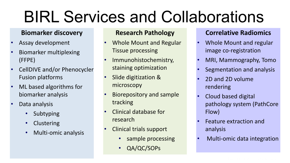
BIRL services and collaborations
Biomarker discovery:
- Assay development
- Biomarker multiplexing (FFPE)
- CellDIVE and/or Phenocycler fusion platforms
- ML based algorithms for biomarker analysis
- Data analysis
- Subtyping
- Clustering
- Multi-omic analysis
Research pathology:
- Whole Mount and regular tissue processing
- Immunohistochemistry, staining optimization, slide digitization and microscopy
- Biorepository and sample tracking
- Clinical database for research
- Clinical trials support:
- Sample processing
- QA/QC/SOPS
Correlative radiomics:
- Whole Mount and regular image co-registration
- MRI, mammography, TOMO
- Segmentation and analysis
- 2D and 2D volume rendering
- Cloud based digital pathology system (PathCore Flow)
- Feature extraction and analysis
- Multi-omic data integration
The lab supports collaborations and offers services in these priority areas:
- biomarker panel development and validation using multiplexing immunofluorescence
- whole-mount tissue processing and large format tissue slide preparation (up to 5”x7”)
- advanced and regular immunohistochemistry for whole-mount and conventional tissue sections
- multimodality image processing and algorithm development, cross-platform image registration and validation
- large capacity, regular and large size tissue slide scanning in bright field and fluorescence modes
- advanced pilot projects and full studies to conduct genomics and transcriptomics profiling research through links with the genomics core facility.
Examples of services:
- Biomarker multiplexing immunofluorescence application and analysis on formalin-fixed paraffin embedded tissue samples and tissue microarrays
- tissue processing and immunohistochemistry, for regular and whole-mount tissue sections (slide sizes 3" x 4" and 5" x 7")
- ultra-rapid tissue-processing protocols for large tissue specimens
- quantitative histological examination of human and preclinical specimens
- antibody staining and optimization
- frozen sections (1" x 3")
- microscopy and digitization of tissue slides (1" x 3" to 5" by 7") in bright field and fluorescence modes
- microcomputed tomography scanning and image analysis
- analysis algorithms, including 2-D and 3-D visualization and correlation tools
- pathological analysis and annotations, immunohistochemistry scoring.
To place an order, download the service request form.


