Equipment
The biomarker imaging research lab is equipped with state-of-the-art research pathology and microscopy instruments. Many of them are available for use by other labs.
Multiplexing immunofluorescence imaging system
A prototype system for research made by GE allows studying co-localization of multiple biomarkers on the same tissue slide.
Tissue processing and staining equipment
These instruments allow fixing and processing of tissue samples by applying conventional and rapid automated microwave protocols, tissue embedding in wax and sectioning of various sizes (from 1”x3” to 5”x7”).
- Pathos automated microwave, Milestone
- Microwave Histos 5 tissue processor, Milestone
- ATP tissue processor, TBS
- Automated immunostainer, Dako
- Microtome SN2500/ultramiller 2600, Leica
- H&E Varistainer, slide agitator
- Automatic immunohistochemistry (IHC) stainer
- Robotic large slide staining system (designed in-house)
- Ventana Discovery instrument for IHC/in situ hybridization provides highest level of protocol flexibility for the most challenging assays and for rapid assay development
- Specimen storage facility and tracking system (for storing slides and tissue blocks)
Digitization and scanners
These microscopes are used to digitize variously sized tissue slides in fluorescent and bright field modes at various resolutions (x1 to x40 and above). Training can be provided to use these instruments.
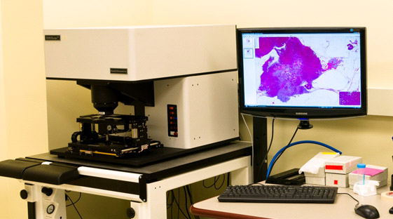
- TissueScope LE
- TissueScope 4000 (2 units)
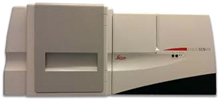
- Leica SCN400
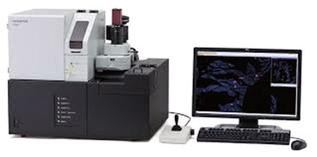
- Olympus VS120


Software and image analysis tools
We operate tissue slide viewing and image analysis software using commercial and open-source viewers. We also have file converters allowing users to view images on various platforms.
- Sedeen Viewer
- ImageJ
- Merkator
- Olympus software
- ClearCanvas, etc.
- Lab-developed algorithms, including titling for large images, vessel counting, ImageJ plug-ins, etc.
MicroCT system
MicroCT specimen imaging system allows CT imaging, volume reconstruction of small samples for the purpose of imaging-pathology correlation. System resolution is 100 um.
Genomics facility
The genomics facility that is part of the biomarker imaging research lab offers in-depth sequencing and many types of molecular analysis. More information about the genomics core facility is available here.


